Transforming Smiles, Restoring Confidence.

What Dental X-Rays Reveal: A Complete Guide to Modern Dental Imaging
image hero:
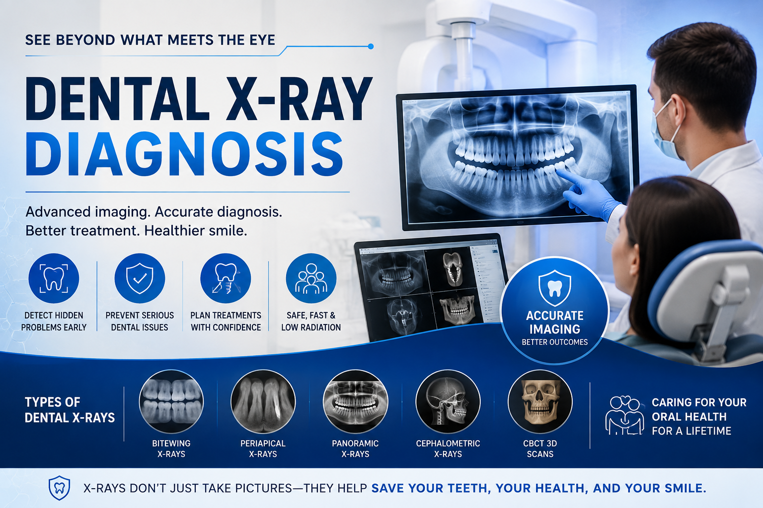
Dental X-rays (radiographs) are among the most valuable diagnostic tools in modern dentistry. While a clinical examination allows your dentist to evaluate the visible surfaces of your teeth and gums, many oral health problems develop beneath the enamel, inside the tooth roots, or within the jawbone—areas that cannot be seen with the naked eye.
Modern digital dental X-rays provide detailed images of your teeth, jawbones, and surrounding structures, allowing dentists to diagnose conditions at their earliest stages. Early detection often means simpler, less invasive, and more affordable treatment while preventing complications that may require extensive restorative procedures later.
Why Dental X-Rays Are Essential
Many dental diseases progress silently. Tooth decay between teeth, infections beneath old fillings, bone loss from gum disease, and impacted wisdom teeth may develop without causing noticeable pain or visible symptoms.
Dental X-rays help your dentist:
-
Detect hidden oral diseases before symptoms appear
-
Confirm findings from a visual dental examination
-
Monitor changes in teeth and jawbone over time
-
Plan restorative and surgical treatments safely
-
Reduce the risk of missed diagnoses
-
Preserve healthy teeth through early intervention
Without radiographic imaging, important conditions may remain undiagnosed until significant damage has already occurred.
What Dental X-Rays Reveal
Hidden Tooth Decay
One of the most common reasons dentists recommend bitewing X-rays is to identify cavities that cannot be seen during a visual examination.
Dental X-rays can reveal:
-
Interproximal cavities developing between adjacent teeth
-
Decay beneath existing fillings
-
Recurrent decay under dental crowns
-
Cavities hidden below the gumline
-
Early enamel demineralization before major tooth destruction occurs
Finding tooth decay early often allows treatment with a small filling instead of requiring a root canal or tooth extraction.
Bone Density Loss from Gum Disease
Periodontal disease gradually destroys the supporting bone around the teeth.
Dental X-rays help dentists evaluate:
-
Loss of jawbone height
-
Horizontal bone loss
-
Vertical bone defects
-
Areas of severe periodontal destruction
-
Progression of periodontal disease over time
These images allow dentists to compare previous X-rays with current ones, helping determine whether gum disease is stable or actively progressing.
Impacted and Un-erupted Teeth
Not every tooth erupts normally.
Dental radiographs can identify:
-
Impacted wisdom teeth
-
Impacted canine teeth
-
Teeth growing sideways
-
Teeth trapped beneath the gums
-
Delayed eruption in children
-
Extra (supernumerary) teeth
Early identification helps prevent crowding, infections, cyst formation, and damage to neighboring teeth.
Dental Infections and Abscesses
An infected tooth does not always cause immediate pain.
-
Dark areas surrounding root tips
-
Bone destruction caused by infection
-
Dental abscesses
-
Dead tooth pulp
-
Chronic infections hidden beneath existing restorations
-
Failed previous root canal treatments
Many infections are discovered during routine examinations before swelling or severe pain develops.
Hidden Cracks, Root Fractures, and Jaw Injuries
Not every fracture is visible during a clinical examination.
Dental imaging can detect:
-
Root fractures
-
Hidden tooth cracks
-
Jawbone fractures
-
Trauma following sports injuries or accidents
-
Structural damage around previously restored teeth
Accurate diagnosis allows dentists to determine whether the tooth can be saved or requires extraction.
Cysts, Tumors, and Other Jaw Abnormalities
Although uncommon, certain pathological conditions develop silently within the jawbone.
Dental X-rays may identify:
-
Fluid-filled cysts
-
Benign jaw lesions
-
Impacted tooth cysts
-
Bone abnormalities
-
Early jaw tumors
-
Changes requiring referral to an oral surgeon
Many of these conditions are discovered incidentally during routine dental examinations before symptoms appear.
Treatment Planning for Complex Dental Procedures
Modern dentistry relies heavily on diagnostic imaging before treatment begins.
-
Bone volume for dental implants
-
Nerve canal location
-
Maxillary sinus position
-
Tooth root anatomy
-
Root canal complexity
-
Orthodontic tooth alignment
-
Wisdom tooth position
-
Jawbone quality
Accurate imaging significantly improves treatment safety and long-term success.
Types of Dental X-Rays
Different types of dental X-rays provide different diagnostic information depending on the clinical situation.
| Dental X-Ray Type | Primary Purpose | Best Used For |
|---|---|---|
| Bitewing X-Ray | Shows the crowns of upper and lower back teeth together. | Detecting cavities between teeth, monitoring existing fillings, and evaluating early bone loss. |
| Periapical X-Ray | Captures the entire tooth from crown to root tip. | Diagnosing root infections, abscesses, root fractures, and surrounding bone conditions. |
| Panoramic X-Ray | Produces a two-dimensional image of the entire mouth, jaws, sinuses, and temporomandibular joints. | Evaluating wisdom teeth, jaw development, impacted teeth, TMJ concerns, and overall oral anatomy. |
| Cone Beam CT (CBCT) | Generates high-resolution three-dimensional images of teeth, bone, nerves, and surrounding structures. | Dental implant planning, complex extractions, oral surgery, endodontics, and advanced diagnostics. |
| Occlusal X-Ray | Captures the entire upper or lower dental arch. | Monitoring children's tooth development, locating impacted teeth, detecting extra teeth, and assessing jaw fractures. |
Digital Dental X-Rays: Safer and More Efficient
Modern dental offices have largely transitioned from traditional film radiographs to digital dental X-rays.
Digital imaging offers several important advantages:
-
Uses approximately 80–90% less radiation than conventional film systems.
-
Produces high-resolution images within seconds.
-
Allows dentists to enlarge and enhance images for more accurate diagnosis.
-
Eliminates the need for chemical film processing.
-
Enables secure electronic storage and easy comparison with previous images.
-
Improves communication by allowing patients to view findings on a chairside monitor.
Because of these benefits, digital radiography has become the standard of care in modern dental practices.
Why Early Detection Matters
Many serious dental conditions begin without pain. By the time symptoms develop, significant damage may already have occurred.
Routine dental X-rays allow dentists to identify hidden disease before it progresses into larger cavities, severe gum disease, abscesses, bone destruction, or tooth loss. Early diagnosis often means more conservative treatment, lower costs, and a better long-term prognosis.
When combined with a comprehensive clinical examination, dental X-rays provide a complete picture of your oral health, enabling your dentist to make accurate diagnoses and develop personalized treatment plans that protect your smile for years to come.
Related Clinical Cases
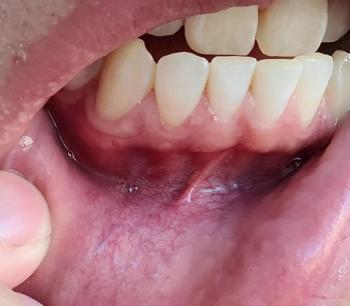
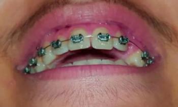
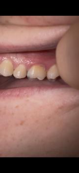
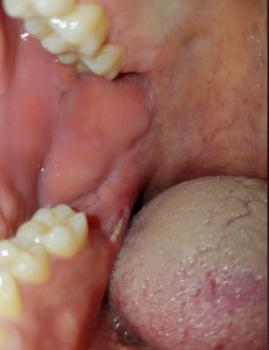
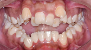
Patients with lower front gum inflammation may also experience tartar buildup, periodontal disease, gum abscesses, or tooth decay. Explore these related clinical case studies.



Other topics:
Other Dental Clinic