Modern Dentistry, Timeless Smiles.

Clinical Case Analysis: Alveolar Bone Loss Following Tooth Extraction
What happens to the jawbone after a tooth is removed? One of the most common concerns patients have before getting a dental implant is whether the jawbone will shrink after tooth extraction.
The answer is yes. When a natural tooth is extracted, the surrounding alveolar bone begins to remodel and gradually lose volume. This process is called alveolar ridge resorption or alveolar ridge atrophy.
Bone loss can affect the width and height of the jawbone and may influence whether a patient needs a bone graft before receiving a dental implant.
Understanding the healing timeline can help patients make informed decisions about tooth replacement.
What Is Alveolar Bone Loss After Tooth Extraction?
Alveolar bone is the part of the jawbone that supports the natural teeth.
A tooth is connected to the surrounding bone through the periodontal ligament. This connection provides mechanical stimulation that helps maintain the supporting bone.
When a tooth is removed, the socket no longer receives the same stimulation. The body begins a natural healing and remodeling process, and the bone surrounding the extraction site may shrink.
This is known as post-extraction alveolar ridge resorption.
Why Does the Jawbone Shrink?
Several biological processes contribute to bone loss:
-
The extraction socket heals after the tooth is removed.
-
The thin bone lining of the socket is remodeled.
-
Bone-forming and bone-resorbing cells reshape the extraction site.
-
The ridge may become narrower and shorter over time.
-
The amount of bone available for a future dental implant may decrease.
This is a normal biological response to tooth removal. It does not necessarily mean that the extraction was performed incorrectly.
1. Post-Extraction Bone Loss Timeline
Bone remodeling begins during the early healing period. The most noticeable changes in ridge dimensions generally occur during the first several months.
Days 1 to 14: Blood Clot and Early Healing
Immediately after tooth extraction, a blood clot forms inside the socket.
The clot is gradually replaced by healing tissue, and the body begins repairing the extraction wound.
During this period:
-
The blood clot helps protect the socket.
-
Inflammatory cells participate in healing.
-
New tissue begins forming.
-
The socket walls start to remodel.
-
The thin bone lining the socket is gradually resorbed and replaced.
Patient question: Can bone loss begin even when the gum looks healed?
Yes. The gum may close over the extraction site while the underlying bone continues to remodel.
Weeks 4 to 12: Early Ridge Remodeling
During the first 2 to 3 months, the extraction site undergoes substantial healing and dimensional changes.
The outer facial bone plate, particularly around front teeth, can be thin and vulnerable to shrinkage.
Possible changes include:
-
Reduction in ridge width.
-
Changes in the contour of the gum and bone.
-
Early loss of the socket's original shape.
-
Decreased bone volume available for implant placement.
The amount of bone loss varies significantly between patients and extraction sites.
Months 3 to 6: Continued Bone Remodeling
Between 3 and 6 months, the extraction site continues to mature.
Some patients experience substantial narrowing of the ridge during this period. The dentist may evaluate the healing site with clinical examination and dental imaging.
Bone loss can be more noticeable when:
-
The facial bone plate was thin.
-
The extraction involved significant trauma.
-
There was existing periodontal disease.
-
The tooth had an infection or bone destruction.
-
No ridge-preservation procedure was performed.
1 Year and Beyond: Long-Term Ridge Atrophy
Bone remodeling may continue after the first year.
The rate of change generally slows over time, but the extraction site can remain smaller than its original dimensions.
Long-term effects may include:
-
A narrower jaw ridge.
-
Reduced support for a removable denture.
-
Increased difficulty placing an implant in the original position.
-
Changes in the gum contour.
-
Additional bone grafting requirements in some cases.
Important: There is no single bone-loss rate that applies to every patient. The commonly quoted percentages and millimeter measurements are averages from particular studies, not a guaranteed prediction for an individual.
2. Horizontal vs. Vertical Bone Loss
Bone loss after tooth extraction can occur in two main directions.
Horizontal Bone Loss
Horizontal bone loss means the ridge becomes narrower from the cheek side toward the tongue or palate.
This is often a major concern for dental implant treatment.
A ridge may retain much of its original height but become too narrow to accommodate an implant safely without additional treatment.
Vertical Bone Loss
Vertical bone loss means a reduction in the height of the supporting ridge.
This can affect implant placement when there is insufficient bone height around the planned implant.
For upper back teeth, the maxillary sinus may also influence available bone height.
Why Does Ridge Width Matter?
A dental implant requires adequate bone volume for stability and proper positioning.
If the ridge is too narrow, the dentist may recommend:
-
Bone grafting.
-
Ridge augmentation.
-
Ridge expansion in selected cases.
-
An alternative implant position.
-
Other treatment options.
The exact procedure depends on the amount and location of bone available.
3. Why Some Extraction Sites Lose More Bone
Not every tooth extraction leads to the same degree of bone loss.
Front Teeth
The upper front teeth often have a thin facial bone plate.
After extraction, this area may experience noticeable changes in the gum and ridge contour.
This is particularly important for patients who want a natural-looking dental implant in the smile zone.
Back Teeth
Molars generally have larger extraction sockets and may involve multiple roots.
The amount of bone loss depends on the anatomy of the site and the condition of the surrounding tissues.
Upper molars may also be affected by maxillary sinus pneumatization, a process in which the sinus expands into the space previously occupied by tooth-supporting bone.
Infection and Bone Damage
A tooth with advanced periodontal disease or a large infection may already have lost supporting bone before extraction.
This means the patient may begin the healing process with less bone than a person who had a healthy tooth removed.
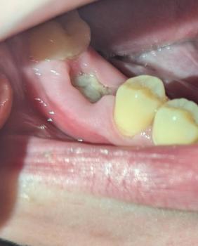
4. Clinical Case Example: Missing Tooth for Several Months
Patient Scenario
A patient had a molar extracted 8 months ago and did not replace the tooth with a dental implant.
The patient now wants an implant but is concerned that the jawbone may have shrunk.
Clinical Considerations
The dentist may evaluate:
-
The width of the remaining ridge.
-
The height of the available bone.
-
The condition of the gum tissue.
-
The location of the inferior alveolar nerve or maxillary sinus.
-
Whether a bone graft is required.
-
The position of the future implant and crown.
Possible Treatment Outcomes
Case A: Adequate Bone Volume
The ridge has sufficient width and height. The dentist may be able to place the implant without additional bone augmentation.
Case B: Narrow Ridge
The ridge has become too narrow for the planned implant. A bone graft may be recommended before or during implant placement.
Case C: Reduced Bone Height
The patient has insufficient bone height, particularly in an upper back tooth area. A sinus lift or another bone augmentation procedure may be considered.
Case D: Existing Infection or Periodontal Disease
The dentist must assess and manage the underlying condition before proceeding with implant treatment.
Clinical Lesson
A missing tooth for 8 months does not automatically mean that a dental implant is impossible.
The decision depends on the actual bone volume, gum condition, and implant treatment plan.
5. How Can You Reduce Bone Loss After Tooth Extraction?
Socket Preservation
Socket preservation is a procedure performed at the time of extraction to help maintain the dimensions of the extraction ridge.
The dentist may place bone-graft material into the socket and use a protective membrane when indicated.
The purpose is to reduce the amount of ridge shrinkage during healing.
Benefits of Socket Preservation
-
May help maintain ridge width.
-
Can improve the conditions for future implant placement.
-
May reduce the need for more extensive augmentation.
-
Helps preserve the shape of the extraction site.
Does socket preservation prevent all bone loss?
No. It helps reduce dimensional changes, but it cannot completely stop natural bone remodeling.
Timely Dental Implant Planning
Patients who plan to replace a missing tooth with an implant should discuss the treatment timeline with their dentist before extraction whenever possible.
Early planning allows the dentist to consider whether socket preservation, immediate implant placement, or another approach is appropriate.
6. When Should You Get a Dental Implant After Extraction?
The timing varies depending on the condition of the extraction site.
| Treatment approach | General timing |
|---|---|
| Immediate implant | Same day as extraction in selected cases |
| Early implant placement | Often around 2–3 months |
| Delayed implant placement | Often 3–6 months or longer |
| Complex bone grafting | May require additional healing time |
These are general treatment categories, not fixed rules.
A dentist may recommend a different timeline depending on the patient's bone healing and clinical findings.
Frequently Asked Questions: Jawbone Loss After Tooth Extraction
1. How quickly does bone loss begin after tooth extraction?
Bone remodeling begins during the early healing period after extraction. The most noticeable dimensional changes often occur during the first few months.
2. Does everyone lose bone after tooth extraction?
Most extraction sites undergo some degree of ridge remodeling and dimensional reduction. The amount varies based on anatomy, healing, infection, bone thickness, and other factors.
3. How much bone do you lose after one year?
Studies have reported different amounts of horizontal and vertical ridge reduction. There is no single number that applies to everyone.
Your dentist can estimate your remaining bone volume using an examination and appropriate imaging.
4. Can I get a dental implant after 1 year of tooth extraction?
Yes. Many patients can receive dental implants one year or more after extraction.
However, some patients may need bone grafting or other preparation if the ridge has become too narrow or too short.
5. Is bone loss after tooth extraction permanent?
Some of the original ridge volume may not return naturally. Bone grafting and other procedures can help rebuild or improve the ridge in selected cases.
6. Can a dental implant stop bone loss?
A dental implant can provide functional stimulation to the surrounding bone and may help maintain bone around the implant.
However, it does not restore every part of the original natural tooth-supporting anatomy. Bone loss can still occur around implants, particularly when peri-implant disease develops.
7. Is bone grafting always necessary before a dental implant?
No. Bone grafting is not necessary for every patient.
The dentist determines whether enough bone is available for safe and stable implant placement.
8. What happens if I wait 5 years after tooth extraction?
The ridge may undergo additional remodeling over time. Some patients may still have adequate bone for an implant, while others may require augmentation.
Waiting does not automatically make implant treatment impossible.
9. Can bone loss change the appearance of my face?
Significant loss of supporting bone and teeth over time may affect the gum contour and, in some cases, facial support.
The extent of any visible change depends on the number of missing teeth, location, and individual anatomy.
10. Can a dentist see bone loss without an X-ray?
A dentist can assess the gum and ridge contour during a clinical examination, but imaging is often needed to evaluate the underlying bone accurately.
Depending on the case, the dentist may recommend a dental X-ray or CBCT scan.
11. Is bone loss worse in the upper front teeth?
The upper front teeth may have thin facial bone plates, making the ridge vulnerable to dimensional changes after extraction.
However, the degree of bone loss varies between patients.
12. Does a tooth infection cause more bone loss?
An infection can damage the supporting tissues and may result in bone loss before extraction.
The dentist must assess the condition of the extraction site and manage infection appropriately.
13. Can I avoid bone grafting if I get an implant immediately?
Immediate implant placement may help reduce the time without a tooth, but it does not guarantee that bone grafting will be unnecessary.
The dentist may still recommend grafting or other procedures depending on the bone defect and implant position.
14. How do I know if I have enough bone for a dental implant?
Your dentist may evaluate:
-
Clinical ridge width.
-
Gum health.
-
Dental X-rays.
-
CBCT imaging when indicated.
-
The planned implant position.
-
The condition of the surrounding teeth.
15. Is it too late to get a dental implant after bone loss?
It is not necessarily too late.
Depending on the severity of bone loss, treatment may include bone grafting, ridge augmentation, or another implant strategy.
Conclusion: Protect Your Jawbone After Tooth Extraction
Alveolar bone loss is a normal part of healing after tooth extraction, but the amount of ridge shrinkage differs from patient to patient.
If you plan to replace a missing tooth with a dental implant, early consultation with a dentist can help determine whether socket preservation or another treatment is appropriate.
The sooner the bone condition is evaluated, the sooner you can understand your available implant options.
Clinical Recommendation
Patients with a missing tooth should discuss their long-term replacement options with a qualified dentist or oral surgeon.
A personalized evaluation is necessary to determine:
-
Whether bone grafting is required.
-
When implant placement should occur.
-
Whether the gum tissue needs treatment.
-
Which implant position is suitable.
-
How long the complete treatment may take.
Medical Disclaimer: This article is for general educational purposes only. It does not replace a professional dental examination, X-ray, or CBCT scan. Dental implant treatment and bone grafting decisions should be made by a qualified dentist or oral surgeon.
Related Clinical Cases
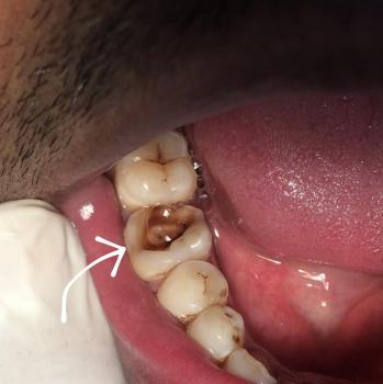
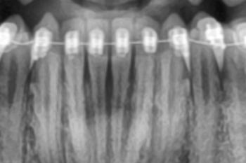
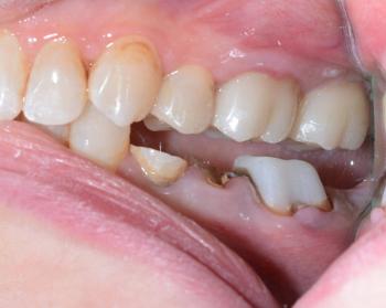
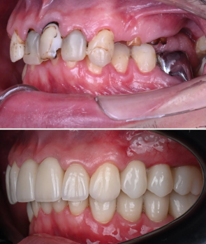
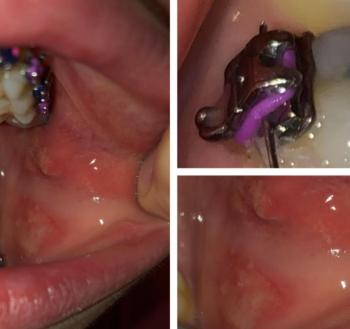
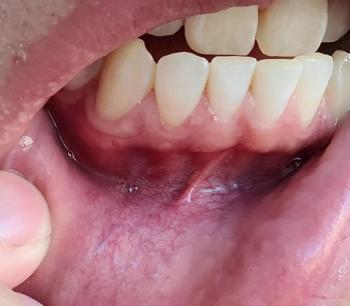
Patients with lower front gum inflammation may also experience tartar buildup, periodontal disease, gum abscesses, or tooth decay. Explore these related clinical case studies.

Other topics:
Other Dental Clinic
 |
 |
 |
 |




