Your Smile, Our Passion.

Back Molar with Deep Fissures Case Analysis
Image:
Severity:
Teeth Problems:
Dental Case Analysis: Back Molar with Deep Grooves and Early Decay Risk
Case Overview
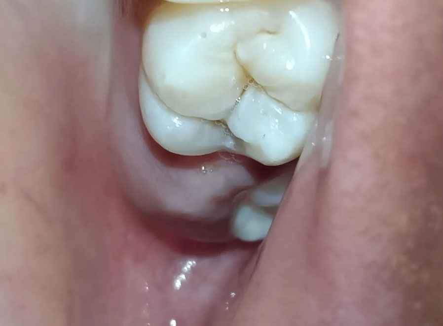
The image shows a posterior molar tooth with deep occlusal grooves (fissures). While the enamel surface appears mostly intact, the depth of the grooves creates a high risk area for early tooth decay due to plaque and food retention.
Zoom 100% Visual Examination
Observed Findings
-
Back molar with pronounced pits and fissures
-
Enamel surface appears smooth but irregular
-
Possible early staining within fissures
-
No obvious fracture or large cavity
-
Saliva pooling around grooves, indicating retention zones
-
Surrounding soft tissue appears normal
-
Adjacent teeth appear intact
Diagnosis
Primary Diagnosis
-
Deep occlusal fissures with high caries risk
Secondary Diagnosis
-
Possible early enamel demineralization
-
Increased susceptibility to future cavities
Treatment Objective
-
Prevent progression to dental caries
-
Seal or protect deep grooves
-
Maintain tooth structure and chewing function
Treatment Process to Execute
Step-by-Step Plan
-
Clinical examination and caries risk assessment
-
Dental X-ray if decay is suspected beneath enamel
-
Professional cleaning of fissures
-
Preventive treatment options:
-
Fissure sealant application
-
Preventive composite restoration if needed
-
-
Oral hygiene instruction and monitoring
Deep Grooves in Back Molars: Early Tooth Decay Risk, Dental Sealants, and Treatment Options
Why Deep Fissures in Back Molars Need Dental Attention
The chewing surfaces of molars naturally contain small pits and grooves called pits and fissures. These anatomical features help the teeth grind food effectively, but deep or narrow grooves can also create areas where plaque and food debris accumulate.
When plaque bacteria break down sugars and fermentable carbohydrates, they produce acids that remove minerals from the tooth enamel. Repeated acid attacks can weaken the enamel and eventually lead to tooth decay. Deep fissures may increase the risk of decay because they can be difficult to clean thoroughly with ordinary brushing.
According to the National Institute of Dental and Craniofacial Research (NIDCR), early tooth decay may develop without noticeable symptoms. A tooth can therefore have an early carious lesion even when the patient does not experience pain. Source: NIDCR — Tooth Decay
Clinical Case Discussion: Back Molar With Deep Fissures
In the referenced case, the back molar is described as having pronounced grooves on its chewing surface, with possible early staining and no obvious large cavity or fracture in the available image.
These findings raise concern about potential plaque retention and early caries. However, deep grooves and dark staining do not automatically mean that a tooth has active decay.
Findings that require careful assessment
-
Deep occlusal fissures: Narrow grooves may retain plaque and food particles, making cleaning more difficult.
-
Staining within the grooves: Brown or dark discoloration may represent harmless staining, arrested decay, or active caries. Color alone cannot establish the diagnosis.
-
Possible early enamel demineralization: The dentist should assess whether the enamel is losing minerals or whether a cavity has already formed.
-
No obvious large cavity: The absence of a visible hole does not exclude early decay or disease extending beneath the enamel.
-
Surrounding gum tissues: The gums should be assessed for redness, swelling, bleeding, and food impaction.
Clinical interpretation: The main concern is the possibility of early occlusal caries in a molar with deep fissures. A definitive diagnosis requires a clinical examination, and dental radiographs may be indicated depending on the findings and suspected extent of disease.
What Happens If Deep Molar Fissures Are Left Untreated?
Deep fissures do not inevitably develop into cavities. However, when active decay develops and the underlying causes remain uncontrolled, the condition can progress through several stages.
Stage 1: Deep fissures without detectable decay
The enamel remains intact, but the grooves may retain plaque. Preventive care, fluoride toothpaste, professional monitoring, and a dental sealant when appropriate can help reduce the risk of decay.
Stage 2: Early enamel demineralization
Repeated acid exposure removes minerals from the enamel. The affected area may develop a chalky white appearance or other discoloration, although early lesions are not always visible to the patient.
At this stage, a dentist may recommend fluoride treatment, dietary changes, plaque control, and monitoring. Selected noncavitated lesions may also be managed with sealants or other minimally invasive treatments.
Stage 3: A cavity develops
If mineral loss continues, the enamel can break down and form a cavity. Once a cavity has formed, fluoride alone generally cannot rebuild the missing tooth structure. The dentist may need to remove damaged tissue and restore the tooth with a filling.
Stage 4: Decay reaches the dentin
Dentin lies beneath the enamel and is less mineralized. As decay advances into dentin, the tooth may become sensitive to sweets, cold, heat, or pressure. Some cavities remain painless despite significant damage.
Treatment depends on the depth and extent of the lesion and may require a larger restoration.
Stage 5: Pulp inflammation or infection
Advanced decay can irritate or infect the dental pulp, which contains nerves and blood vessels. Possible symptoms include spontaneous toothache, lingering sensitivity to temperature, pain that interrupts sleep, or tenderness when biting.
Depending on the pulp diagnosis and whether the tooth can be restored, treatment may involve vital pulp therapy in selected cases, root canal treatment, or extraction if the tooth cannot be predictably saved.
Untreated infection can sometimes progress to an abscess and spread into nearby tissues.
Important: These stages describe a possible progression, not a prediction that every deep fissure will develop into advanced disease.
Treatment Options for Deep Grooves in Molars
The correct treatment depends on whether the tooth is healthy, has early noncavitated decay, contains a cavity, or has deeper structural damage.
1. Dental sealants
A dental sealant is a thin protective material applied to the chewing surface of a tooth. It flows into pits and fissures and creates a barrier that helps prevent plaque and food debris from accumulating in those areas.
Sealants are particularly useful for children and adolescents with newly erupted permanent molars, although selected adults may also benefit based on their cavity risk and the condition of the tooth.
A sealant may also help manage selected noncavitated occlusal caries lesions when the tooth can be adequately assessed and sealed. It is not a substitute for appropriate treatment of a cavity that requires restoration.
2. Fluoride treatment
Fluoride supports enamel remineralization and helps reduce the risk of further mineral loss. A dentist may recommend professional fluoride varnish or other fluoride products according to the patient's age, caries risk, and clinical findings.
Fluoride can help manage early noncavitated lesions, but it cannot replace tooth structure already lost through cavitation.
3. Tooth-colored filling
When decay has produced a cavity, the dentist may remove the affected tooth tissue and restore the defect with a suitable filling material, such as composite resin.
The amount of tooth preparation depends on the lesion's extent and the need to preserve healthy tooth structure.
4. Larger restoration or dental crown
A larger cavity or a molar weakened by substantial structural damage may require a more extensive restoration. A crown may be appropriate when sufficient tooth structure has been lost that a conventional filling would not provide adequate long-term protection.
A crown is not automatically necessary simply because the molar has deep fissures.
5. Root canal treatment
If decay causes irreversible pulp inflammation or pulp necrosis, root canal treatment may be indicated when the tooth is restorable. The procedure involves treating the pulp space and sealing the root canals, followed by an appropriate restoration.
Root canal treatment is not needed for every cavity or every sensitive tooth.
Clinical principle: Choose the least invasive treatment that can predictably control the disease and preserve the tooth, while avoiding unnecessary drilling or restoration.
References: American Dental Association — Dental Sealants and NIDCR — Dental Fillings.
How Dentists Diagnose Decay in Deep Fissures
A dental examination may include:
-
Visual examination: The dentist inspects the grooves for discoloration, enamel breakdown, visible cavities, and other abnormalities.
-
Cleaning and drying: Removing plaque and examining a clean, dry surface can help reveal early changes in the enamel.
-
Clinical assessment: The dentist evaluates the lesion's appearance and activity, the patient's caries risk, symptoms, and previous dental history. Gentle examination may be appropriate; forceful probing that damages an early lesion should be avoided.
-
Dental X-rays when indicated: Bitewing or other appropriate radiographs may help identify decay extending into dentin or areas that cannot be assessed visually. X-rays do not detect every early occlusal lesion.
-
Pulp and structural assessment: If symptoms suggest deeper disease, additional tests may assess pulp status, tenderness, bite-related pain, or possible cracks.
No single visual sign or diagnostic test should be interpreted in isolation.
How to Prevent Tooth Decay in Back Molars
Patients with deep molar grooves can lower their risk of cavities through consistent preventive care.
-
Brush twice daily with fluoride toothpaste, paying particular attention to the chewing surfaces of the back teeth.
-
Clean between the teeth daily using floss or an appropriate interdental cleaner.
-
Reduce frequent consumption of sugary foods and drinks, particularly repeated sipping or snacking throughout the day.
-
Drink water regularly and maintain a balanced diet.
-
Ask a dentist whether fissure sealants or professional fluoride treatment are appropriate.
-
Attend regular dental examinations based on individual risk.
-
Seek assessment for new sensitivity, persistent food trapping, pain when biting, or visible changes in a molar.
Good oral hygiene is important, but deep grooves can remain difficult to clean. Professional prevention and individualized monitoring may therefore be beneficial.
When Should You See a Dentist Urgently?
Arrange a dental appointment promptly if a back molar develops persistent sensitivity, spontaneous pain, a visible cavity, a chipped area, or pain when chewing.
Seek urgent dental care for facial or gum swelling, pus, fever associated with a suspected dental infection, or rapidly worsening pain.
Go to an emergency department immediately if swelling causes difficulty breathing or swallowing, or spreads rapidly into the neck or floor of the mouth. These may indicate a serious spreading infection.
Do not attempt to treat a suspected dental abscess with leftover antibiotics or by placing medication directly into the tooth. Dental infections generally require treatment of the source, and antibiotics are indicated only in selected circumstances.
Expert Dental Conclusion
Deep fissures in back molars are common anatomical features, but their depth and shape can increase the risk of plaque retention and occlusal caries. Early assessment is valuable because a noncavitated lesion may be managed conservatively, whereas an established cavity may require restorative treatment.
For the referenced case, the appropriate next step is to confirm whether the grooves are sound, stained, affected by early demineralization, or associated with a cavity. The dentist can then recommend preventive treatment, a sealant, a filling, or more extensive care according to the actual findings.
The goal is to detect disease early, preserve healthy tooth structure, and prevent avoidable pain and complications.
Healing and Treatment Time Frame
First 14 Days
-
No significant discomfort expected
-
Sealant or restoration stabilizes the surface
-
Normal chewing resumes immediately
After 14 Days
-
Tooth remains protected from decay
-
Low risk of cavity development if hygiene is maintained
If Treatment Is Delayed
Issues That May Scale Up
-
Bacteria penetration into deep grooves
-
Development of occlusal cavity
-
Need for drilling and filling
-
Possible progression to deeper dentin decay
Professional Comments
-
Condition is preventive and manageable
-
Early action avoids invasive treatment
-
Deep fissures are common cavity initiation points
Recommendation
Visit a dental clinic for preventive care and evaluation.
Find a clinic near you:
https://cebudentalimplants.com/map-dental-clinic
Video :
Related Clinical Cases
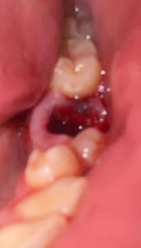
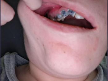
Patients with lower front gum inflammation may also experience tartar buildup, periodontal disease, gum abscesses, or tooth decay. Explore these related clinical case studies.



Other topics:
Other Dental Clinic