Healthy Teeth, Happy Life.

Wisdom Teeth: Impaction, Infection, Cysts & Safe Extraction Planning
image hero:
Country:
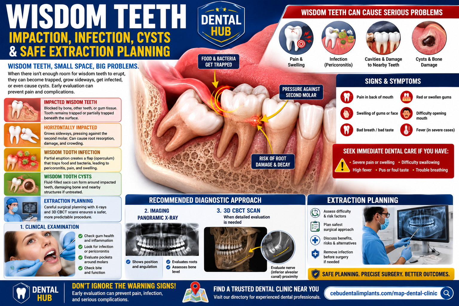
Wisdom teeth—also called third molars—are the final teeth to develop and commonly erupt during the late teenage years or early adulthood. When there is insufficient space in the jaw, a wisdom tooth may become trapped beneath the gum or bone, creating an impacted third molar.
Not every impacted wisdom tooth requires immediate removal. The decision should be based on the tooth's position, symptoms, periodontal condition, damage to neighboring teeth, pathology, and the patient's overall treatment plan. The American Dental Association recommends examination and appropriate X-ray evaluation when wisdom-tooth problems are suspected.
1. What Is an Impacted Wisdom Tooth?
An impacted wisdom tooth cannot erupt into a normal functional position because it is blocked by:
-
Bone
-
Gum tissue
-
The neighboring second molar
-
Insufficient space in the dental arch
-
An abnormal eruption angle
Impaction can be complete, with the tooth remaining entirely under the gum or bone, or partial, where part of the crown becomes visible.
2. Horizontally Impacted Wisdom Teeth
A horizontal impaction occurs when the third molar is positioned sideways rather than vertically.
This can create direct contact with the second molar and may make cleaning extremely difficult. Depending on the position and clinical circumstances, the impacted tooth can be associated with:
-
Food trapping
-
Dental decay
-
Periodontal problems around the second molar
-
Damage to the neighboring tooth
-
Root resorption
-
Recurrent pain or infection
A panoramic radiograph is commonly used to evaluate third-molar position and surrounding structures.
3. Wisdom Tooth Infection: Pericoronitis
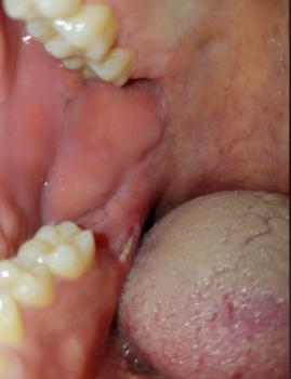
A partially erupted wisdom tooth can leave a flap of gum tissue over part of the crown called an operculum.
Food and bacteria can accumulate underneath this tissue, creating an environment where inflammation and infection can develop.
This condition is known as pericoronitis.
Possible symptoms include:
-
Pain around the back molar
-
Red or swollen gum tissue
-
Bad taste
-
Bad breath
-
Difficulty chewing
-
Swelling around the jaw
-
Difficulty opening the mouth
-
Sometimes fever or more extensive infection
The ADA notes that partially erupted wisdom teeth can allow bacteria to enter the gums and contribute to infection, pain, swelling and jaw stiffness.
4. Can an Impacted Wisdom Tooth Cause a Cyst?
In some cases, a cyst can develop around an impacted third molar.
One example is a dentigerous cyst, which develops around the crown of an unerupted tooth.
A cyst can enlarge within the jaw and potentially affect:
-
Surrounding bone
-
Neighboring teeth
-
Tooth roots
-
The position of nearby structures
Importantly, a cyst cannot be diagnosed from symptoms alone. Radiographic evaluation is required, and definitive diagnosis may require examination of tissue by a qualified clinician.
The ADA recognizes cysts among conditions that may be associated with impacted wisdom teeth and may constitute a reason for removal.
5. When Should a Wisdom Tooth Be Removed?
Extraction may be considered when there is evidence of:
-
Recurrent infection
-
Significant pain
-
Tooth decay that cannot reasonably be restored
-
Gum disease around the wisdom tooth or adjacent second molar
-
Damage to the neighboring tooth
-
Cystic or other pathologic changes
-
A problematic eruption position
-
Other clinically significant disease associated with the tooth
However, impaction alone does not automatically mean extraction is required in every patient. The decision should be individualized.
AAOMS emphasizes clinical and radiographic evaluation and discussion of treatment options before deciding whether an impacted third molar should be removed, exposed, repositioned, or monitored.
6. Diagnostic Process
A comprehensive evaluation usually begins with a clinical examination.
Clinical Examination
The dentist or oral and maxillofacial surgeon may evaluate:
-
Gum inflammation
-
Operculum
-
Pocket depth around the second and third molars
-
Tooth mobility
-
Caries
-
Bite and functional problems
-
Signs of infection
-
Symptoms and previous episodes of pain or swelling
AAOMS specifically emphasizes clinical assessment and periodontal evaluation around third molars and adjacent second molars.
Panoramic X-Ray
A panoramic radiograph can provide a broad view of:
-
Third-molar angulation
-
Depth of impaction
-
Relationship to the second molar
-
Root development
-
Surrounding bone
-
The approximate relationship of lower wisdom-tooth roots to the inferior alveolar canal
Panoramic imaging is commonly used as an initial imaging method when there is a clinical indication to evaluate third molars.
When Is CBCT Useful?
CBCT should not automatically be performed for every wisdom tooth.
Modern ADA recommendations emphasize using dental imaging when clinically justified and using CBCT when lower-exposure imaging cannot provide the information necessary for diagnosis or treatment planning.
For a complex lower wisdom tooth that appears to have a close relationship with the inferior alveolar nerve, the treating clinician may determine that 3D imaging would provide important additional information.
7. Why the Inferior Alveolar Nerve Matters
Lower wisdom-tooth roots can be positioned near the inferior alveolar nerve, which supplies sensation to portions of the lower lip, chin and teeth.
This anatomical relationship is an important consideration when planning extraction.
A panoramic image may suggest whether the roots are close to the nerve canal, but it does not always provide complete three-dimensional information. In selected cases, additional imaging may help the surgeon understand the anatomy and plan the procedure.
The objective is not simply to "get the tooth out." It is to remove a problematic tooth while minimizing avoidable injury to surrounding structures.
8. What Happens During Extraction Planning?
A typical treatment-planning sequence may include:
Step 1 — Clinical examination
Identify symptoms, infection, periodontal problems and the tooth's eruption status.
Step 2 — Appropriate imaging
Use panoramic or other radiographs when clinically indicated.
Step 3 — Assess surgical difficulty
Consider:
-
Tooth angulation
-
Depth
-
Root shape
-
Bone coverage
-
Relationship to the second molar
-
Relationship to nerves and other anatomical structures
-
Patient-specific medical considerations
Step 4 — Discuss treatment options
The clinician should explain the benefits, risks and alternatives, including monitoring when appropriate.
Step 5 — Control active infection when necessary
If significant infection is present, the clinician determines the appropriate way to manage it before or during definitive treatment.
Step 6 — Surgical extraction when indicated
A qualified dentist or oral and maxillofacial surgeon performs the procedure using an appropriate anesthetic and surgical technique.
9. What Happens If You Wait?
An impacted wisdom tooth may remain quiet for years, but that does not mean it can never develop pathology.
Potential problems include:
Food trapping → plaque/bacterial accumulation → gum inflammation → pericoronitis
or
Impaction → contact with second molar → decay/periodontal damage
or
Impacted tooth → cystic/pathologic change → bone or adjacent-tooth damage
Some disease can progress with relatively few symptoms, which is why clinical and radiographic monitoring can be important when a wisdom tooth is retained.
10. When Is It an Urgent Dental Problem?
Seek prompt dental evaluation when there is:
-
Significant swelling
-
Severe wisdom-tooth pain
-
Recurrent infection
-
Facial swelling
-
Fever or feeling systemically unwell
-
Difficulty opening the mouth
-
Difficulty swallowing
-
Rapidly worsening symptoms
Difficulty breathing, severe difficulty swallowing, or rapidly spreading facial/neck swelling requires urgent medical attention.
A serious dental infection should not simply be treated by waiting for symptoms to disappear.
For dental infections, the ADA emphasizes definitive dental treatment when indicated rather than relying on antibiotics alone; antibiotics are generally reserved for situations where systemic involvement or other specific indications are present.
Dental Hub Bottom Line
Wisdom teeth can become much more than a simple "toothache" when they are impacted.
The most important questions are:
Where is the tooth?
Is it damaging the neighboring molar?
Is there infection or periodontal disease?
Is there evidence of cystic or other pathology?
How close are the roots to important anatomical structures?
Is extraction safer than continued monitoring?
A clinical examination plus appropriately selected imaging provides the foundation for answering those questions. Panoramic imaging is commonly used for third-molar assessment, while CBCT is reserved for situations where 3D information is clinically necessary.
Dental Hub educational content is not a substitute for an individualized examination, diagnosis, or treatment plan by a qualified dental professional.
Related Clinical Cases
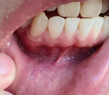
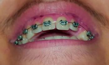
Patients with lower front gum inflammation may also experience tartar buildup, periodontal disease, gum abscesses, or tooth decay. Explore these related clinical case studies.


Other topics:
Other Dental Clinic
 |
 |
 |
 |




