Transforming Smiles, Restoring Confidence.

Recurrent Decay Around Fillings, Hidden Cavities Between Teeth, and Tooth Wear: A Swiss Dentist’s Case Analysis
image hero:
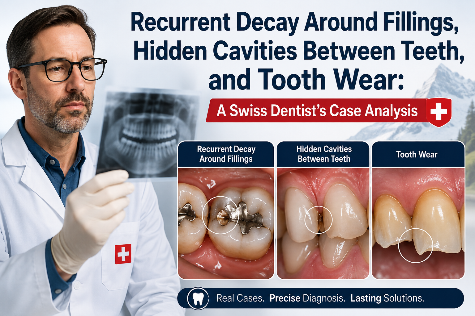
As a dentist working within the Swiss dental environment, I frequently remind patients that a tooth with a filling is not a tooth that can never develop decay again.
Three problems deserve particular attention during routine examinations:
- Recurrent decay around existing fillings
- Hidden cavities between teeth
- Tooth wear and enamel loss
These conditions can be difficult for patients to recognize because they may cause little or no pain during their early stages.
Modern dentistry therefore relies on a combination of clinical examination, risk assessment, visual inspection, appropriate radiographs, photographs, and careful monitoring rather than waiting for a toothache to appear.
Dental caries is a dynamic disease involving demineralization and remineralization, influenced by factors such as plaque biofilm, sugar exposure, saliva, fluoride, and individual risk.
Clinical disclaimer: The case studies in this article are hypothetical educational examples. They do not constitute a diagnosis or individualized treatment recommendation.
1. Recurrent Decay Around Fillings
What Is Recurrent Decay?
Recurrent decay—also called secondary or recurrent caries—refers to caries developing adjacent to or around an existing restoration.
For example, a patient may have a composite filling placed several years ago.
The filling itself may still appear intact, but the tooth structure next to it can develop a new carious lesion.
This is an important distinction:
The filling did not necessarily "get a cavity."
The tooth around the restoration developed a new carious lesion.
The FDI World Dental Federation identifies secondary caries as an important reason restorations are replaced.
Case Analysis 1: Decay Around an Old Filling
Patient
Age: 51
Location: Switzerland
Chief concern: No pain; routine examination
The patient has a large posterior composite restoration that was placed approximately eight years ago.
The patient says:
"The filling feels completely normal."
During examination, I notice a suspicious area along the margin of the restoration.
There is no obvious swelling and no significant sensitivity.
Clinical Examination
I would evaluate:
- Restoration margins
- Plaque accumulation
- Tooth anatomy
- Contact with adjacent teeth
- Signs of cavitation
- Color and texture changes
- Bite relationship
- Symptoms
- Overall caries risk
If the area cannot be adequately evaluated clinically, appropriate radiographic imaging may be considered.
Why Can Decay Develop Around a Filling?
Several factors can contribute.
1. New plaque accumulation
Plaque can accumulate around restoration margins.
2. Frequent sugar exposure
Frequent consumption of fermentable carbohydrates can repeatedly favor acid production and demineralization.
3. Inadequate cleaning
Some restoration margins and interproximal areas are difficult to clean.
4. Changes in saliva
Reduced salivary flow can increase caries risk.
5. Restoration defects
Overhangs, rough surfaces, marginal defects, or other restoration-related problems can contribute to plaque retention.
6. The patient's underlying caries risk
A patient who developed several cavities before receiving fillings can develop new disease after treatment if the underlying risk factors remain active.
The FDI describes caries as a multifactorial disease and emphasizes individualized risk assessment and management rather than focusing exclusively on restoring individual lesions.
Does Every Dark Line Around a Filling Mean Recurrent Decay?
No.
This is a very important point.
A dark margin may represent:
- Staining
- Restoration discoloration
- Surface changes
- Caries
- Shadowing
- Other factors
A dentist should not automatically replace a restoration simply because the margin is darker.
The clinical question is:
Is there evidence that active caries or a clinically significant restoration problem is present?
That requires examination and, when appropriate, additional diagnostic information.
How Is Recurrent Decay Diagnosed?
Depending on the situation, the dentist may use:
- Visual examination
- Dental explorer or other tactile assessment where appropriate
- Intraoral photography
- Bitewing radiographs
- Other radiographic views when indicated
- Digital imaging
- Caries-risk assessment
- Review of previous records
The ADA's current caries-detection guidance emphasizes selecting diagnostic imaging according to the lesion location, anatomy, and clinical findings rather than automatically taking radiographs for every patient.
What Happens If Recurrent Decay Is Found?
Treatment depends on the lesion.
Very early lesion
The dentist may consider preventive or nonrestorative management when appropriate.
Localized cavitated lesion
A minimally invasive restoration may be considered.
Extensive recurrent decay
The existing restoration may need to be repaired or replaced.
Deep decay
The dentist must assess the condition of the pulp and the remaining tooth structure.
Modern caries treatment increasingly emphasizes conservative removal of diseased tissue and preservation of healthy tooth structure when clinically appropriate.
Repair or Replace the Filling?
This is an important clinical decision.
A dentist does not necessarily need to remove an entire restoration every time a localized problem is discovered.
Depending on the situation, options may include:
- Monitoring
- Preventive treatment
- Repairing a localized defect
- Partial replacement
- Complete replacement
The decision depends on:
- Size of the lesion
- Location
- Restoration condition
- Remaining tooth structure
- Caries activity
- Patient risk
- Ability to isolate and clean the area
The objective should be to preserve healthy tooth structure whenever possible.
2. Hidden Cavities Between Teeth
What Are Interproximal Cavities?
Interproximal caries develops on the surfaces between adjacent teeth.
These areas can be difficult to see because the teeth are touching.
A patient can look in the mirror and see apparently healthy teeth while a lesion is developing between them.
This is why professional examination matters.
Case Analysis 2: The Cavity the Patient Could Not See
Patient
Age: 38
Location: Geneva, Switzerland
Chief concern: "My teeth don't hurt."
The patient has good-looking front teeth and no obvious visible cavities.
During examination, however, the dentist notices that the posterior teeth have tightly contacting surfaces that cannot be completely evaluated visually.
Because the patient's clinical history and caries risk justify further assessment, posterior bitewing radiographs are obtained.
The radiograph reveals a suspicious radiolucent area between two posterior teeth.
Why Are Cavities Between Teeth Difficult to Detect?
The problem is anatomical.
The contact point between teeth can hide the lesion.
A dentist may not be able to directly see the entire proximal surface.
The ADA's radiographic recommendations recognize that proximal lesions may require radiographic assessment when the surfaces cannot be adequately examined clinically.
The ADA's newer imaging recommendations likewise emphasize that imaging decisions should be based on the lesion location and clinical circumstances.
What Is a Bitewing X-Ray?
A bitewing radiograph is a dental X-ray designed to capture the crowns of upper and lower posterior teeth and the surrounding bone level.
It is particularly useful for detecting certain cavities between teeth that may not be visible clinically.
But radiographs should not be taken automatically without considering the patient's clinical situation.
The modern approach is:
Clinical examination first → determine the diagnostic question → obtain appropriate imaging when justified.
Can Flossing Prevent Cavities Between Teeth?
Flossing or another appropriate interdental cleaning method can help remove plaque from areas a toothbrush cannot reach.
However, no cleaning technique completely eliminates caries risk.
Other factors remain important:
- Sugar frequency
- Fluoride exposure
- Saliva
- Existing caries
- Plaque control
- Diet
- Individual risk
The FDI recommends fluoridated toothpaste as a key component of caries prevention and advocates twice-daily use at an appropriate concentration and dose.
Can a Cavity Between Teeth Heal?
If the lesion is early and noncavitated, remineralization or arrest may be possible depending on the location and activity.
FDI guidance supports preventive and nonrestorative management of initial carious lesions, with monitoring and fluoride-based approaches.
If a lesion has progressed to significant cavitation and structural breakdown, however, restorative treatment may be necessary.
This is why early detection matters.
3. Tooth Wear and Enamel Loss
Not every defect in a tooth is a cavity.
This is one of the most important distinctions in this case analysis.
Patients sometimes see a flattened tooth, notch, groove, or sensitive area and assume:
"I have a cavity."
But the cause may instead be non-carious tooth wear.
FDI identifies several mechanisms of tooth wear, including:
- Erosion
- Attrition
- Abrasion
- Possible abfraction
These mechanisms may occur independently or together.
What Is Tooth Wear?
Tooth wear means loss of tooth surface structure through processes other than bacterial dental caries.
It may involve:
Erosion
Chemical dissolution caused by acids that are not produced by bacteria.
Attrition
Tooth-to-tooth mechanical wear.
Abrasion
Mechanical wear caused by external objects or forces.
Combined wear
Many patients have more than one mechanism occurring simultaneously.
Case Analysis 3: Progressive Enamel Loss
Patient
Age: 47
Location: Zürich, Switzerland
Chief complaint: "My teeth look shorter."
The patient has no major cavities.
However, the front teeth appear flatter than photographs from several years earlier.
The posterior teeth also show flattened chewing surfaces.
What Would I Investigate?
I would ask about:
- Teeth grinding
- Clenching
- Acidic beverages
- Carbonated drinks
- Citrus consumption
- Frequent acidic foods
- Reflux symptoms
- Vomiting
- Aggressive toothbrushing
- Abrasive products
- Occupational exposure
- Previous dental history
The FDI notes that tooth wear can involve acid exposure, tooth brushing, malocclusion, parafunctional activity, and other mechanical factors. Reduced salivary flow can also influence the process.
Enamel Loss From Acid Erosion
Acid erosion is different from bacterial decay.
The ADA describes dental erosion as a chemical process involving dissolution of dental hard tissue by acids that are not of bacterial origin.
Potential sources include:
Extrinsic acids
- Soft drinks
- Energy drinks
- Acidic fruit juices
- Acidic foods
- Other dietary acids
Intrinsic acids
- Gastric reflux
- Repeated vomiting
Frequent consumption of acidic beverages can be particularly important.
A Typical Erosion Pattern
Imagine a patient who regularly drinks acidic beverages throughout the day.
The patient may say:
"I don't drink much soda at one time."
But the more important issue can be frequency of exposure.
Repeated acid attacks can progressively weaken the tooth surface.
Over time, the enamel may become:
- Smoother
- Thinner
- Flattened
- More translucent
- More susceptible to sensitivity
The ADA identifies frequent consumption of soft drinks, especially carbonated beverages, as an important risk factor for erosive tooth wear.
Tooth Wear From Grinding
Another patient may have a different pattern.
This patient clenches or grinds the teeth.
The dentist may see:
- Flattened cusps
- Matching wear facets
- Chipped enamel
- Fractured restorations
- Muscle tenderness
- Evidence of heavy tooth contact
But tooth wear alone does not prove that grinding is the only cause.
A complete history and examination are necessary.
Tooth Wear Is Often Multifactorial
This is an important clinical principle.
A patient may have:
Acid erosion
Bruxism
Aggressive brushing
Reduced saliva
The resulting tooth wear may be significantly greater than what any single factor would produce.
FDI emphasizes that successful management depends on accurately diagnosing the mechanisms and understanding the contributing factors.
Can Lost Enamel Grow Back?
This is one of the most common questions.
Completely lost enamel does not naturally regenerate.
Mature enamel does not have living cells capable of rebuilding a large amount of lost structure.
However, early mineral loss is different.
Early demineralization can potentially be remineralized under favorable conditions.
This distinction is crucial:
Mineral loss ≠ complete structural loss.
Once significant enamel thickness has physically disappeared, prevention focuses on stopping or slowing further loss and protecting the remaining tooth.
Tooth Sensitivity and Enamel Loss
As enamel becomes thinner, dentin may become more exposed.
Patients may experience:
- Cold sensitivity
- Sweet sensitivity
- Air sensitivity
- Discomfort while brushing
Sensitivity should not automatically be attributed to enamel erosion.
Other possible causes include:
- Gingival recession
- Caries
- Cracked teeth
- Defective restorations
- Pulpal conditions
A dental examination is therefore important.
Comparing the Three Problems
| Condition | Primary Process | Can It Be Hidden? | Typical Diagnostic Approach |
|---|---|---|---|
| Recurrent decay | New caries around restoration | Yes | Clinical exam + imaging when indicated |
| Interproximal decay | Caries between teeth | Very commonly | Clinical exam + bitewing/imaging when appropriate |
| Tooth wear | Non-carious mechanical/chemical loss | Sometimes | Clinical exam + history + photographs |
The treatment is completely different depending on the cause.
Case Review: Three Patients, Three Diagnoses
Patient A — Old Filling
Finding: Suspicious area adjacent to an existing restoration.
Diagnosis to investigate: Recurrent/secondary caries.
Management: Depends on activity, depth, restoration condition, and risk; may range from monitoring to repair or replacement.
Patient B — Hidden Proximal Lesion
Finding: No obvious visible cavity but suspicious interproximal radiographic finding.
Diagnosis to investigate: Interproximal caries.
Management: Depends on whether the lesion is noncavitated, progressing, or cavitated.
Patient C — Shorter Teeth
Finding: Flattened enamel surfaces without obvious caries.
Diagnosis to investigate: Non-carious tooth wear.
Management: Identify and control the underlying causes before considering restorative reconstruction.
Why Diagnosis Comes Before Treatment
One of the most common mistakes in dentistry is treating the appearance rather than the disease process.
Consider a dark area around a filling.
If it is staining rather than active caries, replacing the filling unnecessarily removes healthy tooth structure.
Now consider the opposite:
A patient has an apparently intact restoration, but radiographs reveal a proximal carious lesion.
Simply looking at the filling may miss the disease.
And consider tooth wear.
If a dentist simply places crowns on worn teeth without identifying the underlying cause, the new restorations may also experience excessive wear.
Therefore:
Diagnose the cause before rebuilding the tooth.
Modern Swiss Dental Approach: Prevention and Minimal Intervention
A contemporary approach to these conditions is increasingly focused on:
Early detection
→ Risk assessment
→ Disease control
→ Minimal intervention
→ Monitoring
The FDI supports a shift toward prevention and arrest of caries lesions, with operative treatment reserved for situations where it is actually indicated.
The ADA's restorative caries guideline similarly supports conservative approaches for many moderate and advanced carious lesions.
This philosophy is particularly valuable when treating patients over many decades.
Every millimeter of healthy tooth structure matters.
Frequently Asked Questions
Can decay come back under a filling?
Yes. New caries can develop adjacent to or around an existing restoration. This is commonly referred to as recurrent or secondary caries.
Does a filling last forever?
No. Restorations have finite service lives and can eventually develop defects, fracture, wear, staining, or recurrent caries around them.
Does every old filling need to be replaced?
No. A filling that remains clinically satisfactory does not automatically need replacement simply because it is old.
Why does decay develop around my filling?
Possible contributors include plaque retention, frequent sugar exposure, inadequate cleaning, restoration defects, reduced saliva, and the patient's overall caries risk.
Can a dentist see every cavity without an X-ray?
No. Some cavities, particularly between teeth, may not be directly visible during a routine examination.
Why are cavities between teeth called "hidden cavities"?
Because adjacent teeth can obscure the affected surfaces, making direct visual detection difficult.
Can flossing remove a cavity between my teeth?
No. Flossing can remove plaque and help prevent disease, but it cannot physically remove an established cavity.
Can an early cavity between teeth heal?
Some early, noncavitated lesions can potentially be arrested or remineralized depending on their activity and location. Preventive treatment and monitoring are important.
Does tooth wear mean I have cavities?
No. Tooth wear and dental caries are different processes.
What causes enamel loss?
Possible causes include acid erosion, tooth-to-tooth wear, abrasion, parafunctional activity, and combinations of these factors.
Can enamel grow back?
Significant lost enamel does not naturally regenerate. Early mineral loss, however, may be remineralized under appropriate conditions.
Can soda cause enamel loss?
Frequent exposure to acidic soft drinks can contribute to erosive tooth wear.
Does grinding wear away enamel?
Grinding and other parafunctional forces can contribute to tooth wear, although wear usually requires assessment of all possible contributing factors.
Is tooth wear reversible?
The lost tooth structure itself generally cannot be naturally regenerated. The important goal is to identify and control the cause before additional structure is lost.
Should worn teeth automatically receive crowns?
No. The cause, severity, symptoms, remaining tooth structure, function, aesthetics, and long-term risks should be evaluated first.
Can acid erosion and cavities happen at the same time?
Yes. A patient can have both caries and erosive tooth wear.
How can I protect my enamel?
Important measures may include:
- Twice-daily brushing with fluoridated toothpaste
- Cleaning between teeth
- Limiting frequent sugar exposure
- Reducing frequent acidic drinks
- Maintaining adequate saliva
- Addressing reflux or other relevant medical factors
- Regular dental examinations
FDI recommends fluoridated toothpaste as a core caries-prevention strategy.
When Should You See a Dentist?
Arrange an examination if you notice:
- A filling that feels different
- A broken filling
- Food repeatedly getting trapped around a restoration
- New tooth sensitivity
- A dark area near a filling
- Teeth becoming visibly shorter
- Chipped or flattened teeth
- Persistent sensitivity to cold
- Pain when biting
- A gap developing between teeth
- Bleeding or swelling around a tooth
Do not wait for severe pain before seeking evaluation.
Final Clinical Perspective
From a Swiss dental perspective, I would summarize these three conditions with one principle:
Not every abnormal-looking tooth needs a filling, and not every cavity is visible to the patient.
A filling can remain healthy for years, but new decay can develop around it.
A tooth can look completely normal while a lesion is developing between two tightly contacting teeth.
And teeth can lose significant enamel without any bacterial cavity being present.
These conditions require different diagnostic strategies.
Recurrent decay
Look carefully around existing restorations and evaluate the patient's overall caries risk.
Hidden interproximal cavities
Use clinical examination and appropriate radiographic imaging when the surfaces cannot be adequately evaluated directly.
Tooth wear and enamel loss
Determine whether acid, tooth-to-tooth contact, abrasion, parafunction, reduced saliva, or a combination of factors is responsible.
The modern approach is not simply:
"Find a hole → drill → fill."
It is:
Detect → Diagnose → Identify the cause → Prevent progression → Preserve healthy tooth structure → Restore only when necessary → Monitor over time.
That approach allows us to protect natural teeth for as long as possible while avoiding unnecessary treatment.
And in long-term dentistry, that is often the most important goal of all.
Dental Hub clinical note: Diagnosis of recurrent caries, interproximal lesions, and tooth wear requires an individualized examination. Radiographs should be prescribed according to clinical need, and treatment should be based on the specific tooth, lesion activity, caries risk, symptoms, and overall oral condition.
Related Clinical Cases
Patients with lower front gum inflammation may also experience tartar buildup, periodontal disease, gum abscesses, or tooth decay. Explore these related clinical case studies.


Other topics:
Other Dental Clinic