Strong Teeth, Fresh Breath, Brighter You.

Simulated U.S. Dental Case: Acute Pericoronitis From an Impacted Mandibular Wisdom Tooth
Country:
1. Simulated Case Presentation
Patient Profile
A 22-year-old patient presents to an outpatient oral surgery clinic in the United States with a 3-day history of progressively worsening pain in the lower-right posterior jaw. The patient reports localized swelling, discomfort while chewing, and increasing difficulty opening the mouth.
Subjective Symptoms
The patient describes:
-
Throbbing pain radiating toward the right ear and temple
-
Localized swelling around the lower-right wisdom tooth
-
Foul taste and intermittent metallic or purulent discharge
-
Pain when chewing
-
Mild restriction in mouth opening (trismus)
-
Low-grade fever
Clinical Examination
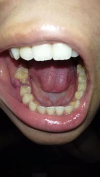
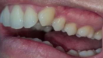
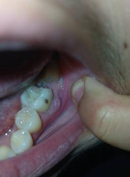
Intraoral examination reveals a partially erupted mandibular right third molar (#32) with an inflamed soft-tissue operculum covering part of the crown.
Clinical findings include:
-
Erythematous, edematous opercular tissue
-
Purulent material expressed from the opercular pocket
-
Localized tenderness
-
Difficulty accessing and cleaning the area
-
Tender right submandibular lymph nodes
-
Temperature of approximately 38.1°C (100.6°F)
The combination of local infection, fever, lymphadenopathy, and trismus suggests that the inflammatory process is more significant than uncomplicated localized gum irritation.
Radiographic Findings
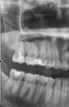
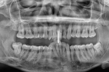
A panoramic radiograph is obtained, with CBCT considered when additional three-dimensional anatomical information is necessary.
Imaging demonstrates:
-
Mesioangular impaction of tooth #32
-
Contact or close proximity between #32 and the distal aspect of the mandibular second molar (#31)
-
A deep plaque-retentive periodontal area between the two teeth
-
Early localized crestal bone loss
-
No obvious acute cystic lesion
The relationship between the impacted third molar and #31 is clinically important because plaque retention and periodontal inflammation can affect the adjacent second molar.
2. Clinical Assessment & Diagnosis
Primary Diagnosis
Acute pericoronitis associated with an impacted mandibular third molar.
Pericoronitis occurs when the soft tissue surrounding a partially erupted wisdom tooth becomes inflamed or infected. The operculum can trap bacteria, food debris, and plaque, creating an environment favorable to recurrent inflammation.
Secondary Consideration
The clinician should also evaluate for a localized periodontal infection or abscess involving the third-molar region, particularly when purulent drainage and periodontal pocketing are present.
Because this simulated patient has fever, lymphadenopathy, and trismus, the clinician must also assess whether the infection remains localized or is beginning to spread into adjacent fascial spaces.
3. Standard U.S. Treatment Pathway
Treatment should be individualized following an examination, appropriate imaging, medical-history review, and assessment of infection severity.
Phase I: Control the Acute Infection
The immediate objective is to control the acute inflammatory process, relieve symptoms, and determine whether the infection can be safely managed in an outpatient setting.
1. Local Irrigation and Debridement
The dentist or oral surgeon may gently irrigate and debride the area beneath the operculum to remove:
-
Food debris
-
Plaque
-
Necrotic material
-
Purulent material
Local treatment is an important component of managing pericoronitis.
Antiseptic rinses may be recommended when clinically appropriate. Chlorhexidine may be prescribed in selected cases, although treatment should be determined by the treating dental professional.
2. Antibiotics When Systemic Involvement Is Present
Antibiotics are not routinely required for every case of pericoronitis.
They become more appropriate when there are signs suggesting systemic involvement or spreading infection, such as:
-
Fever
-
Malaise
-
Significant swelling
-
Lymphadenopathy
-
Trismus
-
Cellulitis
-
Evidence of spreading infection
In this simulated case, the presence of fever, tender lymph nodes, purulent drainage, and trismus would prompt the treating clinician to consider systemic antimicrobial therapy.
The specific antibiotic, dose, duration, and need for allergy precautions should be determined by the treating clinician based on the patient's medical history, allergies, local resistance patterns, and current dental prescribing guidance.
3. Pain Control
Pain management may include an NSAID such as ibuprofen when medically appropriate. Acetaminophen may also be used alone or in combination with an NSAID when appropriate.
Patients should follow the dosage instructions provided by their clinician and avoid NSAIDs or acetaminophen when contraindicated by their medical history, medications, allergies, or other factors.
4. Reassessment Before Definitive Surgery
The patient should be reassessed after the acute phase has been addressed.
The clinician evaluates:
-
Pain and swelling
-
Fever
-
Mouth opening
-
Drainage
-
Lymph-node tenderness
-
Ability to eat and drink
-
Evidence of spreading infection
-
Condition of the adjacent second molar
The timing of definitive extraction is individualized. The tooth does not necessarily need to remain untreated for a fixed 7–14-day period. In some circumstances, extraction during the acute episode may be appropriate, while in others the infection is first stabilized before surgery.
The decision depends on the severity of infection, anatomical complexity, access, anesthesia considerations, and the surgeon's clinical judgment.
5. Definitive Surgical Management
Once the acute condition has been appropriately evaluated and stabilized, the long-term treatment for a problematic impacted third molar is frequently surgical extraction.
Extraction may be recommended when the tooth is associated with:
-
Recurrent pericoronitis
-
Significant periodontal disease
-
Dental caries
-
Damage to the second molar
-
Repeated infection
-
Cystic or other pathological changes
-
A position that creates a substantial risk of future disease
Surgical Approach
For a partially or fully impacted mandibular wisdom tooth, an oral and maxillofacial surgeon may:
-
Administer appropriate local anesthesia.
-
Provide sedation when clinically indicated.
-
Reflect the gum tissue to access the tooth.
-
Remove a limited amount of surrounding bone when necessary.
-
Section the tooth into smaller pieces if required.
-
Remove the tooth.
-
Irrigate the surgical site.
-
Place sutures when indicated.
-
Provide postoperative instructions.
The exact procedure varies according to the depth and orientation of the tooth and its relationship to the inferior alveolar and lingual nerves.
6. What About the Opposing Upper Wisdom Tooth?
The original clinical scenario proposes removing the opposing maxillary third molar at the same time.
This is not automatically required.
An opposing wisdom tooth may be considered for extraction when it is:
-
Over-erupted
-
Traumatizing the inflamed operculum
-
Difficult to maintain
-
Carious
-
Periodontally compromised
-
Otherwise associated with disease or significant future risk
The decision should be based on the actual position and condition of the opposing tooth rather than routinely removing it simply because the lower wisdom tooth is being extracted.
7. Anesthesia and Sedation in the U.S.
Wisdom-tooth surgery can be performed using different anesthesia approaches.
Local Anesthesia
Local anesthesia is commonly used and completely numbs the surgical area while the patient remains conscious.
Nitrous Oxide Sedation
Nitrous oxide may be used to reduce anxiety and improve patient comfort.
IV Sedation
Intravenous sedation can provide deeper relaxation and is commonly used for patients undergoing multiple or surgically difficult extractions.
General Anesthesia
General anesthesia is a deeper level of anesthesia and is reserved for selected situations based on the patient's needs, procedure complexity, medical history, and facility capabilities.
An oral and maxillofacial surgeon determines the safest anesthesia approach after evaluating the patient.
8. When the Case Becomes a Medical Emergency
Most pericoronitis cases can be managed in a dental setting, but certain symptoms indicate possible progression into a serious deep-space infection.
Emergency Warning Signs
Immediate emergency evaluation is appropriate when a patient develops:
-
Rapidly increasing facial or neck swelling
-
Difficulty breathing
-
Difficulty swallowing
-
Drooling
-
Significant inability to open the mouth
-
Swelling extending toward the floor of the mouth or neck
-
Altered voice
-
Severe systemic illness
-
Rapid clinical deterioration
A suspected deep neck-space infection can threaten the airway and may require hospital treatment, intravenous antibiotics, surgical drainage, and airway management.
Ludwig's angina is one example of a potentially life-threatening spreading infection involving the floor of the mouth and submandibular spaces.
In such circumstances, the priority is airway and systemic infection management—not routine outpatient wisdom-tooth extraction.
9. U.S. Insurance and Financial Considerations
The cost of treatment depends on the patient's insurance plan, geographic location, provider, surgical complexity, anesthesia, imaging, and whether treatment occurs in a dental office or hospital.
Insurance considerations can include:
-
Dental-network status
-
Annual deductible
-
Annual maximum
-
Surgical extraction benefits
-
Sedation coverage
-
Radiographic imaging benefits
-
Waiting periods
-
Preauthorization requirements
-
Coordination between dental and medical insurance
Emergency evaluation and definitive surgical treatment may be billed differently depending on the clinical circumstances and insurance plan.
Patients should request a written treatment estimate and ask the dental or oral-surgery office to verify benefits before elective surgery whenever practical.
10. Clinical Takeaway
This simulated case represents a symptomatic, infected mandibular third molar with acute pericoronitis and signs of systemic involvement.
The important clinical sequence is:
Recognize the infection → assess severity → provide local treatment and appropriate systemic management when indicated → monitor for spreading infection → reassess → determine definitive treatment.
The presence of fever, lymphadenopathy, purulent drainage, or trismus should prompt careful evaluation rather than treating the condition as simple gum irritation.
For a repeatedly problematic impacted wisdom tooth, definitive extraction may prevent recurrent episodes and reduce the risk of further damage to the adjacent second molar. However, the timing and surgical approach must be individualized by the treating dentist or oral and maxillofacial surgeon.
This simulated case is for educational purposes only. Antibiotic selection, dosing, anesthesia, extraction timing, and emergency management must be determined by a licensed clinician after evaluating the individual patient.
Related Clinical Cases
Patients with lower front gum inflammation may also experience tartar buildup, periodontal disease, gum abscesses, or tooth decay. Explore these related clinical case studies.
Other topics:
Other Dental Clinic