From Pain to Perfect — We Care for Your Smile.

Clinical Case Review: Periapical (Radicular) Cysts of the Jaw
From a German dental perspective, a periapical—or radicular—cyst is an important inflammatory lesion associated with a diseased or non-vital tooth. These lesions develop within the tissues surrounding the root apex and are generally connected to longstanding pulpal infection and apical inflammation.
However, there is an important clinical distinction: a radiolucency at the root apex cannot automatically be called a cyst. Periapical granuloma, abscess, cyst, scar tissue, and even some non-inflammatory jaw lesions can have overlapping radiographic appearances. Histopathologic examination remains the definitive method for distinguishing several of these entities.
1. Understanding the Clinical Case
A typical clinical scenario may involve a patient who has:
-
Deep untreated dental caries
-
Previous dental trauma
-
Extensive restoration
-
Previous root canal treatment
-
Recurrent infection
-
A tooth that has become necrotic
Bacteria and their by-products can enter the root canal system and trigger inflammation around the root apex. Persistent inflammation can lead to destruction of the surrounding periodontal tissues and alveolar bone.
The American Association of Endodontists notes that apical periodontitis is the most frequently encountered periapical pathological process and that a periapical radiolucency can represent several different pathological conditions.
The important clinical question
When an X-ray shows a dark area around a tooth root, the dentist should not immediately conclude:
"This is definitely a cyst."
Instead, the clinician should establish a differential diagnosis based on the patient's history, pulp status, clinical examination, imaging, and—when indicated—histopathology.
2. How a Patient May Present
Periapical cysts can remain clinically silent for a considerable period.
A patient may have:
-
No pain
-
No swelling
-
No obvious tooth symptoms
-
A previously traumatized tooth
-
A heavily restored tooth
-
An asymptomatic non-vital tooth
The lesion may therefore be discovered incidentally during a routine periapical or panoramic radiographic examination.
As the lesion becomes larger, however, it may produce:
-
Localized swelling
-
Expansion of the cortical bone
-
Facial asymmetry
-
Tooth displacement
-
Tooth mobility
-
Pressure symptoms
-
Recurrent infection
-
Drainage through a sinus tract
A large lesion may also become clinically significant because of its relationship to the maxillary sinus, nasal cavity, or mandibular nerve canal.
3. Diagnostic Protocol in Modern Dental Practice
Step 1: Medical and Dental History
The dentist should first establish the history of the tooth.
Important questions include:
-
Has the tooth previously been painful?
-
Was there dental trauma?
-
Has the tooth undergone root canal treatment?
-
Is there a large restoration?
-
Has the patient experienced swelling?
-
Has there been drainage?
-
Has the tooth changed color?
-
Has previous treatment failed?
The medical history is equally important because systemic conditions and previous cancer history can alter the differential diagnosis and the need for biopsy.
4. Pulp and Periapical Testing
A suspected periapical lesion should be evaluated together with the involved tooth.
Depending on the clinical situation, testing can include:
-
Cold testing
-
Heat testing
-
Electric pulp testing
-
Percussion
-
Palpation
-
Periodontal probing
-
Mobility assessment
Pulp testing should not be interpreted in isolation. The AAE emphasizes integrating pulp testing with medical and dental history, clinical examination, and radiographic findings.
The central clinical question is:
Is the tooth actually necrotic, and does the lesion anatomically and biologically fit an endodontic origin?
5. Conventional Dental Imaging
Periapical radiographs are often the first imaging method used to evaluate suspected apical disease.
A lesion may appear as a:
-
Well-defined radiolucency
-
Corticated radiolucency
-
Larger periapical bone defect
-
Lesion associated with a non-vital tooth
But radiographic appearance alone cannot reliably differentiate a cyst from a granuloma or other pathology.
The AAE specifically emphasizes that several periapical pathological processes can be radiographically indistinguishable and that histology is the gold standard when definitive tissue diagnosis is required.
6. Where CBCT Adds Clinical Value
Cone-beam computed tomography can provide three-dimensional information when conventional imaging does not adequately answer the clinical question.
CBCT may help demonstrate:
-
True lesion dimensions
-
Cortical expansion
-
Cortical perforation
-
Root anatomy
-
Relationship to adjacent roots
-
Inferior alveolar nerve canal
-
Maxillary sinus floor
-
Other anatomical structures
Modern guidance recognizes CBCT as an established imaging modality when appropriately indicated and correctly interpreted.
Important limitation
CBCT can show where the lesion is and how extensive it is, but it does not automatically tell the dentist what the lesion is histologically.
A large radiolucency is not automatically a cyst.
7. Histopathology: The Diagnostic Confirmation
This is one of the most important lessons in this case review.
A periapical cyst has a characteristic histological structure, including an epithelial lining and connective-tissue wall.
However, other inflammatory and non-inflammatory lesions can mimic it clinically and radiographically.
The AAE recommends biopsy in situations including persistent pathology, lesions inconsistent with endodontic disease, suspicious clinical findings, and when sufficient tissue is obtained during periapical surgery.
Therefore:
Radiograph = suspicion
Clinical examination + testing = diagnostic direction
Histopathology = definitive tissue diagnosis when indicated
8. Treatment Strategy: Preserve the Natural Tooth When Possible
The primary objective of modern endodontic treatment is to eliminate the infection while preserving a functional natural tooth whenever this is predictably achievable.
For an endodontically treatable tooth associated with apical disease, nonsurgical root canal treatment or retreatment may be appropriate.
Treatment generally involves:
-
Accessing the root canal system
-
Mechanical cleaning and shaping
-
Appropriate irrigation
-
Disinfection
-
Obturation
-
Coronal restoration
-
Clinical and radiographic follow-up
Contemporary endodontic guidelines emphasize appropriate diagnosis, aseptic treatment, irrigation, root canal preparation and filling, followed by reassessment.
9. Can a Periapical Cyst Heal After Root Canal Treatment?
This question requires nuance.
Some lesions diagnosed clinically as cystic may decrease or resolve after successful nonsurgical endodontic treatment because the underlying microbial stimulus has been eliminated.
However, a true cystic lesion is a histopathological diagnosis, and not every cyst-like radiolucency behaves identically.
The AAE notes that true cystic lesions are among the situations in which surgical management may ultimately be necessary when appropriate nonsurgical treatment does not resolve the pathology.
Therefore, I would not advise a patient to judge treatment success simply by whether the lesion looked smaller on one early radiograph.
Healing must be evaluated over time.
10. When Surgical Treatment Becomes Relevant
Surgery may be considered when:
-
Nonsurgical endodontic treatment has failed
-
Retreatment is not feasible or predictable
-
Persistent periapical disease remains
-
A true cystic lesion is suspected or confirmed
-
There is a persistent extraradicular problem
-
A biopsy is required
-
An anatomical or procedural problem prevents adequate nonsurgical treatment
Depending on the case, treatment may involve apical microsurgery, lesion removal, and management of the root end.
11. Apical Microsurgery and Enucleation
A surgical approach may involve:
-
Local anesthetic administration
-
Reflection of a mucoperiosteal flap
-
Access to the periapical lesion
-
Removal of pathological tissue
-
Root-end resection when indicated
-
Root-end preparation
-
Placement of a suitable root-end filling material
-
Closure and postoperative monitoring
The removed tissue may be submitted for histopathological examination.
In selected modern microsurgical cases, materials such as bioceramic root-end filling materials can be used.
Recent clinical literature also describes combinations of apical surgery, cyst enucleation, bioceramic materials, bone grafting, and platelet concentrates in carefully selected cases.
12. Decompression of Large Cystic Lesions
A particularly interesting treatment option for selected large cystic periapical lesions is decompression.
The concept is to reduce pressure within the lesion and encourage gradual reduction in size.
A decompression technique may involve placement of a drain or tube that permits continued drainage.
This approach can potentially reduce the size of a large lesion before definitive surgery and may help protect important anatomical structures.
A 2026 AAE clinical review describes decompression as a potentially useful option for selected large cystic periapical lesions and emphasizes that lesion size alone does not prove that a lesion is a cyst.
That distinction is extremely important.
13. Bone Regeneration After Treatment
After removal or decompression of a large periapical lesion, the resulting bony defect may gradually fill with new bone.
In selected surgical cases, clinicians may consider regenerative materials such as:
-
Bone graft materials
-
Barrier membranes
-
Platelet concentrates
-
Advanced platelet-rich fibrin
-
Other regenerative biomaterials
These materials should not be presented as universally necessary. Their use depends on the size and anatomy of the defect, surgical objectives, patient factors, and clinician judgment.
14. Prognosis and Follow-Up
The prognosis depends on considerably more than simply the presence of a cyst.
Important factors include:
-
Correct diagnosis
-
Quality of root canal treatment
-
Coronal seal
-
Tooth restorability
-
Presence of persistent infection
-
Root anatomy
-
Surgical technique when required
-
Patient-specific factors
-
Lesion characteristics
A large radiolucency may take considerable time to remodel after successful treatment.
Follow-up may include clinical examination and repeat periapical imaging. In selected cases, CBCT can provide additional information when conventional imaging is insufficient.
The objective is to document:
symptom resolution → reduction of the lesion → progressive bone healing → stable long-term outcome.
15. What I Would Tell a Patient in My Dental Chair
If your dentist tells you:
"There is a cyst at the end of your tooth,"
do not immediately assume that you have a dangerous tumor or that your tooth must be extracted.
First, ask:
-
Is the tooth vital or non-vital?
-
Does the lesion appear to originate from an endodontic infection?
-
Do I need a CBCT?
-
Is nonsurgical root canal treatment appropriate?
-
Does the lesion require referral to an endodontist or oral surgeon?
-
Should tissue be submitted for histopathological examination?
-
How will healing be monitored?
These questions turn a frightening X-ray finding into a structured diagnostic and treatment plan.
Clinical Bottom Line
From a German dentist's evidence-based perspective, the most important message is this:
A periapical radiolucency is a finding—not automatically a final diagnosis.
The clinician must connect the radiographic appearance with pulp testing, clinical findings, patient history, and appropriate imaging. When tissue is removed or the lesion behaves atypically or persists despite appropriate treatment, histopathological evaluation becomes particularly important.
Modern endodontic care increasingly combines precise diagnosis, minimally invasive tooth-preserving treatment, microsurgical techniques, three-dimensional imaging when indicated, and long-term monitoring.
For the patient, the ultimate goal is straightforward:
eliminate the infection, preserve the natural tooth whenever predictably possible, restore the surrounding bone, and make sure the lesion heals.
Clinical Disclaimer
This Dental Hub article is an educational review and does not diagnose or prescribe treatment for an individual patient. A suspected periapical cyst requires examination by a qualified dentist, endodontist, oral and maxillofacial surgeon, or other appropriate dental specialist. Treatment should be based on the patient's individual clinical findings, imaging, medical history, and—when indicated—histopathological diagnosis.
Related Clinical Cases
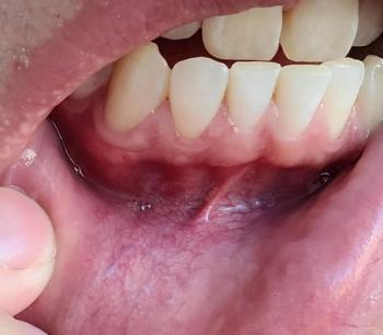
Patients with lower front gum inflammation may also experience tartar buildup, periodontal disease, gum abscesses, or tooth decay. Explore these related clinical case studies.


Other topics:
Other Dental Clinic