We Bring Back the Sparkle in Your Smile.

Jaw Cysts and Tumors: A USA Dentist’s Clinical & Research Review
A jaw cyst or tumor can develop quietly inside the maxilla (upper jaw) or mandible (lower jaw), sometimes without pain or obvious changes to the mouth. Some lesions are small and relatively straightforward to manage, while others can expand, displace teeth, weaken bone, affect nerves, or require complex oral and maxillofacial surgery.
From a U.S. dental perspective, the key clinical principle is simple: an abnormal area on an X-ray is not automatically a diagnosis. Clinical examination, dental imaging, and—when indicated—biopsy and microscopic examination must be considered together. Different lesions can look remarkably similar on radiographs while requiring very different treatments.
1. Jaw Cysts vs. Jaw Tumors
What is a jaw cyst?
A cyst is generally a pathological cavity that may contain fluid, keratin, inflammatory material, or other contents and may have an epithelial lining depending on its classification.
Many jaw cysts are odontogenic, meaning they originate from tissues or cellular remnants associated with tooth development.
Common examples include:
-
Radicular/periapical cyst
-
Dentigerous cyst
-
Odontogenic keratocyst (OKC)
-
Glandular odontogenic cyst
-
Lateral periodontal cyst
-
Other developmental and inflammatory cysts
Odontogenic cysts may be discovered during routine dental imaging because many produce few or no symptoms during their early stages.
What is a jaw tumor?
A tumor is a proliferation of tissue that may be benign or malignant.
Odontogenic tumors arise from tissues involved in tooth formation, while other jaw tumors and bone lesions can originate from different cellular components of the maxilla or mandible.
Importantly, benign does not necessarily mean harmless. Some benign jaw tumors can be locally aggressive and destroy surrounding bone.
2. Important Jaw Cysts and Tumors
Radicular Cyst
A radicular cyst is associated with an inflammatory process arising from a non-vital tooth, commonly following longstanding pulpal infection.
A patient may have no significant symptoms until the lesion becomes large or secondarily infected.
Clinical evaluation should include assessment of the involved tooth's vitality and the surrounding bone.
Dentigerous Cyst
A dentigerous cyst is associated with the crown of an unerupted tooth.
It is frequently encountered around impacted teeth, particularly mandibular third molars, although other teeth can be involved.
Small lesions may be discovered incidentally. Larger lesions can cause expansion, tooth displacement, or other structural changes.
Odontogenic Keratocyst
The odontogenic keratocyst (OKC) is particularly important because it can behave more aggressively than many conventional jaw cysts and has a recognized tendency toward recurrence.
Historically, it was called a "keratocystic odontogenic tumor," but modern classification uses odontogenic keratocyst as the preferred terminology.
OKCs may occur in the posterior mandible and can extend considerably within cancellous bone before producing obvious external swelling.
Multiple OKCs can occur in association with Gorlin syndrome, making the patient's medical and family history clinically important.
Ameloblastoma
Ameloblastoma is a benign odontogenic tumor, but it can be locally aggressive.
It commonly affects the mandible and may produce progressive expansion, tooth displacement, root resorption, and significant destruction of surrounding bone.
Radiographically, some lesions can resemble other cystic or radiolucent processes. Histopathology is therefore critical when the diagnosis is uncertain.
Treatment is individualized according to tumor type, size, location, and biological behavior.
Odontoma
Odontomas contain dental hard tissues such as enamel and dentin and are generally considered benign odontogenic tumors.
They are often discovered because a permanent tooth fails to erupt normally.
Imaging may reveal a characteristic radiopaque mass associated with an unerupted tooth.
Central Giant Cell Lesions
Central giant cell lesions occur within the jawbone and may range from relatively slow-growing lesions to more aggressive processes.
They can produce radiolucent changes on imaging and may cause expansion or tooth displacement.
Because several jaw lesions can have overlapping radiographic appearances, the diagnosis must be established through appropriate clinical, imaging, and pathological evaluation.
3. What Symptoms Should Patients Watch For?
Many jaw cysts and tumors are initially asymptomatic.
A lesion may be discovered unexpectedly during a panoramic X-ray or other dental imaging obtained for an unrelated dental problem.
As a lesion enlarges, possible findings include:
-
Jaw or facial swelling
-
Facial asymmetry
-
Tooth displacement
-
Loosening of teeth
-
Changes in the bite
-
Delayed tooth eruption
-
Expansion of the jawbone
-
Chewing discomfort
-
Difficulty opening the mouth
-
Drainage or recurrent infection
-
Persistent sinus tract
-
Numbness or altered sensation
Numbness deserves particular attention. A lesion affecting or compressing a nerve pathway can produce altered sensation in the lip, chin, cheek, or other areas.
Persistent swelling, unexplained bone changes, progressive tooth displacement, or unexplained numbness should be evaluated rather than simply observed without professional guidance.
4. How a U.S. Dental Team Investigates a Jaw Lesion
A proper diagnosis is usually a step-by-step process.
Step 1: Clinical Examination
The dentist or oral surgeon evaluates:
-
Facial symmetry
-
Intraoral swelling
-
Tooth mobility
-
Tooth vitality
-
Periodontal condition
-
Occlusion
-
Soft tissues
-
Neurologic symptoms
-
Medical and dental history
Tooth vitality testing is particularly important when evaluating suspected odontogenic cysts because it helps distinguish lesions associated with diseased or non-vital teeth from other conditions.
Step 2: Panoramic and Periapical Imaging
A panoramic radiograph provides a broad view of the jaws and can reveal:
-
Radiolucent areas
-
Radiopaque lesions
-
Impacted teeth
-
Root displacement
-
Bone expansion
-
Relationship to adjacent structures
Periapical imaging can provide more detailed information about individual teeth and their surrounding bone.
However, a radiographic appearance alone does not always establish the final diagnosis.
Different lesions can have overlapping appearances.
5. Where CBCT Fits Into Modern Dental Diagnosis
Cone-beam computed tomography, or CBCT, provides three-dimensional information about the bony anatomy.
Depending on the clinical situation, CBCT can help clinicians evaluate:
-
Lesion dimensions
-
Cortical expansion
-
Cortical perforation
-
Relationship to tooth roots
-
Inferior alveolar nerve canal
-
Maxillary sinus
-
Nasal cavity
-
Other important anatomical structures
The major advantage is spatial information that cannot be obtained from a conventional two-dimensional radiograph.
However, CBCT should not be viewed as a substitute for pathology. It is primarily an imaging tool for evaluating the bony lesion and its anatomical relationships.
6. Why Biopsy and Histopathology Matter
This is one of the most important points in evaluating jaw lesions.
Imaging can suggest a diagnosis, but pathology may be necessary to establish it.
Some lesions can appear remarkably similar radiographically. For example, certain dentigerous cysts, odontogenic keratocysts, and cystic ameloblastomas may overlap in their imaging characteristics while requiring substantially different management.
Depending on the circumstances, an oral and maxillofacial surgeon may perform:
Incisional biopsy
A representative portion of the lesion is removed for microscopic examination.
Excisional biopsy
The entire lesion is removed when appropriate, often when it is relatively small and can safely be treated in one procedure.
The pathology report is then interpreted together with the patient's clinical and radiographic findings.
7. Treatment: Why There Is No Single "Jaw Cyst Treatment"
Treatment depends on:
-
Exact diagnosis
-
Lesion size
-
Location
-
Biological behavior
-
Relationship to teeth
-
Relationship to nerves and other structures
-
Cortical involvement
-
Recurrence risk
-
Patient-specific medical considerations
Enucleation
Enucleation involves removing the cystic lesion from the surrounding bone.
It can be appropriate for selected cystic lesions, particularly when the lesion is well defined and complete removal can be performed safely.
Decompression or Marsupialization
For selected large cystic lesions, a surgeon may choose decompression or marsupialization.
The objective is to reduce internal pressure and allow the lesion to decrease in size over time.
This can sometimes make subsequent definitive surgery less extensive and may help protect nearby teeth or anatomical structures.
Resection
More aggressive lesions may require removal of a larger portion of the affected jaw.
This is particularly relevant for certain aggressive tumors where recurrence risk and local destruction must be carefully considered.
Treatment decisions should be individualized rather than based solely on the lesion's appearance on one X-ray.
8. Jaw Reconstruction After Major Surgery
When substantial bone is removed, reconstruction may be necessary to restore:
-
Jaw continuity
-
Facial contour
-
Chewing function
-
Speech
-
Oral function
-
Future dental rehabilitation
Depending on the case, reconstruction can involve bone grafting or microvascular free-tissue reconstruction.
In selected patients, dental implants may eventually be considered as part of functional rehabilitation.
The timing of implant placement depends on healing, reconstruction stability, pathology, radiation exposure when applicable, and the overall treatment plan.
9. Complications of Untreated or Aggressive Lesions
A growing jaw lesion can progressively weaken the bone.
Potential complications include:
-
Significant bone destruction
-
Tooth displacement
-
Root resorption
-
Infection
-
Nerve involvement
-
Facial asymmetry
-
Difficulty chewing
-
Pathological fracture in severely weakened bone
-
Recurrence after treatment
Large lesions deserve particular attention because the risk of structural complications can increase as the lesion expands.
10. Recurrence and Long-Term Follow-Up
Treatment does not always mark the end of care.
Certain lesions have a meaningful recurrence risk, particularly some aggressive odontogenic lesions.
For these patients, long-term clinical and radiographic surveillance may be recommended.
The appropriate follow-up interval depends on the pathology, treatment method, surgical margins, recurrence history, and individual patient factors.
A lesion that has been removed successfully still needs appropriate follow-up when its biology warrants it.
11. The Clinical Takeaway From a U.S. Dentist's Perspective
When a dentist discovers an unusual radiolucency, radiopacity, or mixed lesion in the jaw, the correct response is not to assume that it is simply a cyst.
The diagnostic process should ask:
Where is the lesion?
Is it associated with a tooth?
Is the tooth vital?
What does the panoramic image show?
Does CBCT add important three-dimensional information?
Is the lesion expanding or destroying cortical bone?
Are nerves or other anatomical structures involved?
Does the lesion require biopsy?
What does histopathology show?
This integrated approach is essential because radiographic appearances can overlap among different jaw lesions, and the final diagnosis can substantially change the treatment plan.
Dental Hub Patient Message
Do not panic if your dentist discovers a "cyst" or "lesion" on an X-ray.
Many jaw lesions are benign, and some are discovered incidentally before they cause significant symptoms. But an unexplained lesion should be properly evaluated.
If you have persistent jaw swelling, unexplained tooth movement, delayed eruption, recurrent drainage, facial asymmetry, or numbness, arrange a professional dental evaluation.
Depending on the findings, your dentist may refer you to an oral and maxillofacial surgeon or an oral and maxillofacial pathology service for further investigation.
Important terminology update
The term "keratocystic odontogenic tumor (KCOT)" is historical. The lesion is now generally referred to as an odontogenic keratocyst (OKC) in contemporary classification.
Clinical disclaimer
This Dental Hub review is for education and does not diagnose an individual patient. A jaw cyst, tumor, or other bone lesion can only be diagnosed through an appropriate clinical evaluation, imaging, and, when indicated, histopathologic examination.
Research basis: Contemporary reviews emphasize that jaw lesions require integration of clinical findings, radiographic characteristics, and histology, because different entities can have overlapping presentations.
Related Clinical Cases
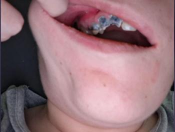
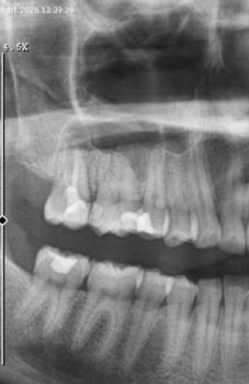
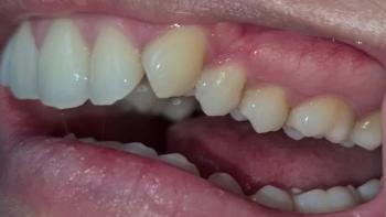
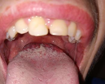
Patients with lower front gum inflammation may also experience tartar buildup, periodontal disease, gum abscesses, or tooth decay. Explore these related clinical case studies.



Other topics:
Other Dental Clinic
 |
 |
 |
 |




