Your Smile, Perfected with Precision.

Bone Loss After Tooth Extraction: Causes, Timeline, Treatment & Prevention
Clinical Overview: Alveolar Ridge Resorption
Alveolar bone resorption is a normal physiological response to tooth extraction. Once the periodontal ligament (PDL) is removed, the lack of functional stimulation triggers bone remodeling, with increased osteoclastic activity and progressive loss of ridge volume.
What Happens After Extraction?
- First 3–6 months: The greatest dimensional changes generally occur during this period.
- Horizontal bone loss: The extraction ridge may lose several millimeters of width, with greater reduction commonly occurring on the facial/buccal aspect.
- Vertical bone loss: Ridge height can also decrease, particularly where the facial bone is thin.
- Long-term changes: Remodeling continues at a slower rate after the initial healing period.
- Functional consequences: Reduced ridge volume can make future implant placement, prosthetic design, and soft-tissue aesthetics more challenging.
Important: The amount of bone loss varies considerably between patients and depends on factors such as socket anatomy, periodontal health, infection, extraction technique, smoking, systemic health, and whether ridge-preservation treatment is performed.
Why Does Bone Loss Occur?
The periodontal ligament normally provides mechanical stimulation to the surrounding alveolar bone. After extraction, this stimulation disappears.
The healing process includes:
Tooth extraction → blood clot → inflammatory/healing phase → woven bone formation → remodeling → reduction of ridge dimensions
The buccal/facial plate is particularly vulnerable because it is often thinner than the lingual or palatal plate.
Clinical Consequences
Progressive ridge resorption can result in:
- Narrower alveolar ridges
- Reduced vertical bone height
- Buccal or facial contour collapse
- Changes in gingival architecture
- Less favorable implant positioning
- Greater difficulty achieving ideal emergence profiles
- Increased need for future bone augmentation
- Complications with removable prosthetic stability
In the maxilla, the residual ridge commonly remodels in a superior and posterior direction. In the mandible, progressive resorption can reduce the vertical height of the residual ridge and increase the clinical significance of the inferior alveolar canal.
United States Clinical Practice Context
Management of post-extraction bone loss in the United States is generally based on evidence-based dental and implant-treatment principles. Organizations such as the American Dental Association, American Academy of Periodontology, and American Association of Oral and Maxillofacial Surgeons provide professional guidance relevant to extraction, periodontal care, and implant therapy.
Alveolar Ridge Preservation
When future implant placement or other restorative treatment is anticipated, alveolar ridge preservation (ARP) may be considered at the time of extraction.
Common approaches include:
- Particulate bone graft materials
- Allografts or xenografts
- Collagen plugs or membranes
- Resorbable barrier membranes
- Primary or secondary soft-tissue closure depending on the clinical situation
The goal is not necessarily to prevent all post-extraction remodeling, but to reduce dimensional collapse and preserve a more favorable ridge architecture.
Implant Treatment
Depending on the clinical circumstances, treatment may involve:
- Immediate implant placement
- Early implant placement
- Delayed implant placement
- Socket preservation
- Guided bone regeneration (GBR)
- Sinus augmentation
- Horizontal or vertical ridge augmentation
Treatment should be individualized according to the available bone, soft-tissue anatomy, infection status, esthetic demands, and patient's restorative goals.
Comprehensive Clinical Assessment
1. Pre-Extraction Risk Assessment
Before removing a tooth, clinicians should evaluate factors that may influence subsequent ridge remodeling.
Periodontal Phenotype
Thin facial bone and thin soft-tissue phenotypes generally present greater risk of contour collapse following extraction.
Existing Pathology
Consider:
- Periodontal destruction
- Periapical pathology
- Endodontic failure
- Root fracture
- Acute infection
- Existing facial-plate defects
Extensive pre-existing bone destruction can reduce the amount of intact socket architecture available for preservation.
Extraction Technique
Atraumatic extraction is important because fracture of the facial/buccal plate can increase the severity of the resulting defect.
Techniques may include:
- Periotomes
- Root sectioning
- Controlled luxation
- Specialized extraction systems
- Piezosurgery in selected cases
2. Diagnostic Imaging
CBCT Evaluation
Cone-beam computed tomography (CBCT) can provide three-dimensional information when conventional radiographs are insufficient for treatment planning.
It may help evaluate:
- Facial and lingual bone thickness
- Residual ridge dimensions
- Root anatomy
- Maxillary sinus location
- Inferior alveolar canal
- Existing bone defects
- Implant positioning requirements
CBCT should be used when the additional three-dimensional information is clinically justified rather than automatically for every extraction.
3. Treatment Planning Decision Matrix
| Clinical Scenario | Potential Treatment Approach | Primary Objective |
|---|---|---|
| Intact socket, limited restorative risk | Socket preservation when indicated | Reduce ridge collapse and maintain future restorative options |
| Thin facial/buccal plate | Ridge preservation or guided bone regeneration | Maintain facial contour and bone volume |
| Compromised socket wall | Particulate graft + barrier membrane when indicated | Reconstruct deficient ridge architecture |
| Severe horizontal defect | Horizontal ridge augmentation | Increase ridge width for appropriate implant positioning |
| Severe vertical defect | Advanced augmentation techniques | Restore adequate vertical bone dimensions |
| Immediate implant candidate | Immediate implant ± grafting | Maintain architecture while shortening treatment time in selected cases |
No single grafting technique is appropriate for every extraction socket. The treatment decision should be based on the anatomy, infection status, restorative plan, esthetic requirements, and clinician assessment.
4. Long-Term Prognostic Factors
Successful preservation of the ridge does not end with graft placement or implant surgery.
Prosthetic Loading
Implants should be positioned and restored to provide appropriate biomechanical loading and minimize unfavorable stress concentrations.
Plaque Control
Excellent oral hygiene is essential for maintaining periodontal and peri-implant health.
Periodontal Maintenance
Regular professional maintenance allows clinicians to monitor:
- Gingival inflammation
- Periodontal pocketing
- Implant stability
- Peri-implant tissue health
- Crestal bone changes
- Prosthetic complications
Soft-Tissue Stability
Adequate soft-tissue architecture and appropriate implant positioning can contribute to long-term esthetic and functional stability.
Key Takeaway
Bone loss after tooth extraction is a normal biological process, but the extent of ridge remodeling can vary substantially. The greatest dimensional changes usually occur during the early healing period. Careful extraction technique, appropriate risk assessment, selective ridge-preservation procedures, and three-dimensional treatment planning can help maintain bone volume and improve future restorative options.
Clinical note: This information is for educational purposes and does not replace an individualized examination, radiographic assessment, or treatment plan from a qualified dental professional.
Related Clinical Cases
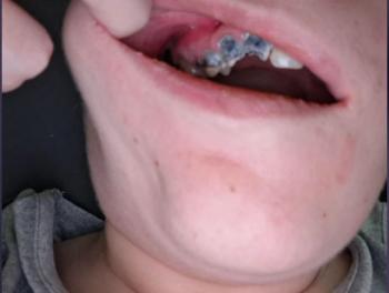
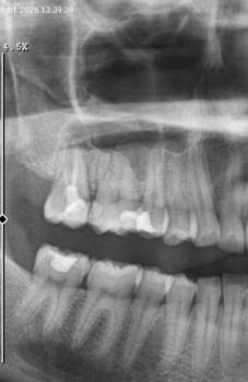
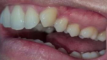
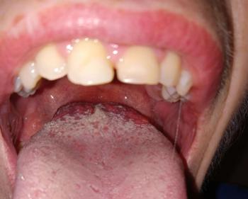
Patients with lower front gum inflammation may also experience tartar buildup, periodontal disease, gum abscesses, or tooth decay. Explore these related clinical case studies.



Other topics:
Other Dental Clinic
 |
 |
 |
 |




