Healthy Teeth, Happy Life.

Lateral Tongue Lesion Case Analysis and Healing Timeline
Image:

Severity:
Teeth Problems:
Clinical Guidance: Lateral Tongue Lesion, Red Patches, and White Areas
Understanding a Lesion on the Side of the Tongue
A lateral tongue lesion is an abnormal area affecting the side or border of the tongue. It may appear as a red patch, white area, ulcer, thickened surface, or painful sore. Some lesions develop after accidental biting, repeated friction against a sharp tooth, or irritation from dental appliances.
However, not every red or white tongue lesion is caused by trauma. Oral candidiasis, inflammatory conditions, persistent ulcers, and potentially malignant disorders may produce overlapping appearances. An in-person examination is necessary to distinguish among these possibilities.
The side of the tongue is an important area to examine carefully because persistent lesions in this location can occasionally indicate oral cancer or a precancerous condition.
Possible Causes of Red and White Patches on the Lateral Tongue
1. Traumatic tongue injury
Accidental biting, sharp tooth edges, broken fillings, and repeated friction can irritate the tongue's delicate surface. A traumatic ulcer may cause redness, tenderness, and a shallow sore. Healing generally begins after the source of irritation is removed.
2. Oral candidiasis (thrush)
This fungal infection can cause white patches, redness, soreness, or a burning sensation. Risk factors include recent antibiotic use, inhaled corticosteroids, dry mouth, dentures, and certain medical conditions. White patches may sometimes wipe away, leaving a red surface, but this feature alone cannot establish the diagnosis.
Antifungal treatment should be based on a clinician's assessment rather than the appearance of a photograph alone.
3. Aphthous ulcers
Canker sores are usually shallow, painful ulcers with a pale or yellowish center and a red border. They can occur on the tongue and other movable oral tissues. A typical canker sore heals within one to two weeks, but persistent or atypical ulcers require professional assessment.
4. Leukoplakia and other potentially malignant disorders
A persistent white patch that cannot be explained by another condition may require assessment for leukoplakia. Red patches and mixed red-and-white lesions also deserve attention. Some potentially malignant lesions have few or no symptoms, so the absence of pain does not guarantee that a lesion is harmless.
5. Oral cancer
Oral cancer can sometimes appear as a non-healing ulcer, persistent red or white patch, firm lump, thickened area, or unexplained bleeding. Additional warning signs include tongue numbness, difficulty moving the tongue, trouble swallowing, or a lump in the neck.
These symptoms do not automatically mean cancer is present. Nevertheless, suspicious lesions need prompt examination and, when indicated, biopsy.
Clinical guidance from the American Dental Association emphasizes a comprehensive oral examination and biopsy or specialist referral for suspicious lesions.
How Long Should a Tongue Lesion Take to Heal?
A minor traumatic lesion may improve within several days after the irritating factor is removed. Many uncomplicated traumatic ulcers heal within approximately 7–14 days, although healing varies according to the cause and severity.
| Observation | Recommended action |
|---|---|
| Small sore after accidental biting that is steadily improving | Protect the area and monitor healing |
| Redness or discomfort that persists without clear improvement | Arrange a dental or medical examination |
| Ulcer, red patch, or white patch lasting two weeks or longer | Seek prompt professional assessment |
| Firm lump, unexplained bleeding, progressive enlargement, or tongue numbness | Arrange urgent evaluation; do not wait for the lesion to become painful |
| Difficulty breathing or inability to swallow saliva | Seek emergency medical care |
A two-week threshold is a practical safety guideline, not a reason to delay care for a suspicious or worsening lesion. A clinician may recommend earlier investigation depending on the appearance and history.
Recommended Clinical Examination
A dentist, oral medicine specialist, or oral and maxillofacial surgeon may assess:
-
Location and size: Whether the lesion affects the side, underside, or tip of the tongue.
-
Surface characteristics: Whether the area is ulcerated, red, white, thickened, irregular, or bleeding.
-
Consistency: Whether there is unusual firmness or a lump beneath the surface.
-
Possible sources of irritation: Sharp teeth, fractured restorations, biting habits, or dental appliances.
-
Medical and social history: Recent medications, tobacco use, alcohol exposure, immune status, and the duration of symptoms.
-
Associated findings: Enlarged neck lymph nodes, difficulty swallowing, numbness, or changes in tongue movement.
If the lesion appears suspicious or does not resolve as expected, a biopsy with histopathological examination may be necessary. A biopsy is the standard method for confirming or excluding many important tissue diagnoses, including oral cancer.
Photographs, visual screening devices, and adjunctive tests cannot reliably replace a clinically indicated biopsy.
Safe Home Care While Waiting for Assessment
For a lesion that appears consistent with minor irritation, the following measures may help reduce discomfort:
-
Rinse gently with warm salt water and spit it out. Avoid excessively salty solutions.
-
Choose soft foods and avoid very hot, spicy, acidic, or rough foods that aggravate the sore.
-
Drink adequate water, particularly if the mouth feels dry.
-
Brush gently with a soft-bristled toothbrush and maintain routine oral hygiene.
-
Avoid smoking, tobacco products, and alcohol while the tissue heals.
-
Do not scrape, cut, burn, or peel the lesion.
-
Arrange dental care if a sharp tooth or damaged restoration repeatedly rubs against the tongue.
Do not use leftover antibiotics or antifungal medication without appropriate assessment. Antibiotics do not treat fungal infections, and unnecessary medication can cause adverse effects.
When to Seek Specialist Care
A persistent lateral tongue lesion should not be managed indefinitely with home remedies. Prompt evaluation is especially important if it grows, becomes firm, bleeds without an obvious cause, develops an irregular ulcerated surface, or is accompanied by numbness or a neck lump.
If a dentist suspects a potentially malignant disorder, referral to an oral medicine specialist, oral and maxillofacial surgeon, or an appropriate head-and-neck specialist may be needed.
Clinical takeaway: Many tongue lesions are benign and heal after irritation is removed. The safest approach is to identify the cause, monitor expected healing, and investigate lesions that persist, progress, or have suspicious features.
Frequently Asked Questions About Lateral Tongue Lesions
1. What causes a red and white patch on the side of the tongue?
Possible causes include accidental biting, friction from sharp teeth, oral candidiasis, inflammatory conditions, and potentially malignant disorders. A clinical examination is needed because these conditions can look similar.
2. Is a lateral tongue lesion always a sign of oral cancer?
No. Many lesions are caused by minor trauma or other benign conditions. However, persistent red or white patches, non-healing ulcers, firm lumps, and unexplained bleeding need professional assessment to exclude serious disease.
3. How long does a traumatic tongue ulcer take to heal?
Many uncomplicated traumatic ulcers improve within several days and heal within about 7–14 days after the cause of irritation is removed. If the lesion persists for two weeks, worsens, or has suspicious features, arrange an examination.
4. Can a sharp tooth cause a lesion on the side of the tongue?
Yes. A sharp tooth edge, fractured filling, or repeated biting can injure the lateral tongue. A dentist can identify and address the source of trauma. Do not attempt to file a tooth yourself.
5. Do white patches on the tongue mean oral thrush?
Not necessarily. White patches can result from several conditions, including candidiasis, frictional changes, and leukoplakia. A clinician can assess whether the patch can be removed, examine the underlying tissue, and determine whether further testing is required.
6. Should I scrape off a white tongue lesion?
No. Avoid scraping, peeling, or cutting a localized white patch. This can injure the tissue and may delay appropriate assessment. A persistent or unexplained patch should be examined professionally.
7. Can saltwater rinses cure a lateral tongue lesion?
Saltwater rinses may soothe minor irritation and help keep the mouth comfortable, but they do not treat every underlying cause. They cannot cure oral cancer or reliably treat candidiasis. Persistent lesions still require examination.
8. Does a painless tongue lesion need attention?
Yes. Some important oral lesions are painless, especially in their early stages. A painless ulcer, lump, or red or white patch that persists should not be ignored.
9. When is a tongue lesion biopsy necessary?
A clinician may recommend biopsy when a lesion looks suspicious, remains unexplained, or fails to resolve as expected. The decision depends on the clinical findings and history. A negative adjunctive screening test should not delay biopsy or referral when a lesion remains concerning.
10. Which specialist should examine a persistent tongue lesion?
Start with a licensed dentist or physician. Depending on the findings, you may be referred to an oral medicine specialist, oral and maxillofacial surgeon, oral pathologist, or ear, nose, and throat (ENT) specialist.
11. Can I use antibiotics for a tongue ulcer?
Not unless a qualified clinician identifies a reason to prescribe them. Many ulcers are not caused by bacterial infections. Unnecessary antibiotics can cause side effects and contribute to antimicrobial resistance.
12. When is a tongue lesion an emergency?
Seek emergency medical care if tongue swelling causes difficulty breathing or swallowing saliva. Rapidly increasing swelling, especially with fever or spreading facial or neck swelling, also warrants urgent assessment.
13. Can a photograph confirm the diagnosis of a tongue lesion?
No. A photograph can help document a lesion's appearance and changes over time, but it cannot reliably establish the diagnosis or assess tissue firmness. An in-person examination and, in some cases, biopsy are necessary.
14. Can smoking or alcohol increase concern about a persistent tongue lesion?
Yes. Tobacco use and alcohol consumption are established risk factors for oral cancer, particularly when exposures occur together. Avoiding tobacco and limiting or avoiding alcohol are important preventive measures, but people without these risk factors can also develop oral cancer.
15. What should I do if the lesion improves but does not disappear completely?
Arrange a dental or medical review. Partial improvement does not establish the cause or exclude a serious condition. The clinician can decide whether continued observation, treatment, referral, or biopsy is appropriate.
Medical Disclaimer
This article provides general educational information and is not a substitute for an individual diagnosis or treatment plan. A tongue lesion cannot be definitively diagnosed from a photograph or online description alone. Seek prompt professional evaluation for persistent, worsening, unexplained, or suspicious oral lesions.
Teeth Case – Full Analysis and Provisional Diagnosis (Zoom 100%)
Medical disclaimer:
This assessment is based solely on the provided image. A definitive diagnosis requires an in-person oral examination. Tongue lesions that persist, enlarge, or become painful must be evaluated by a licensed dental or medical professional.
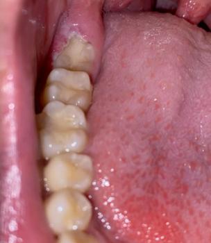
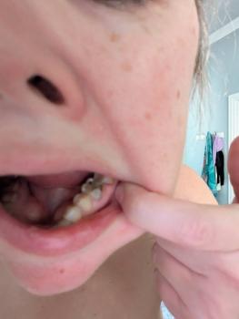
1. Visual Findings (Deep Examination)
-
Tongue (lateral/side surface):
-
Irregular reddish inflamed area with scattered white patches on the lateral border.
-
Appearance suggests surface irritation with secondary coating, not a thick fixed plaque.
-
-
Adjacent tissues:
-
Mild redness of surrounding mucosa.
-
No obvious heavy swelling in the image.
-
-
Possible contributing factors:
-
Repeated tongue friction against teeth
-
Accidental biting
-
Fungal overgrowth in an irritated area
-
Dry mouth or recent antibiotic use
-
2. Most Likely Diagnosis (Provisional)
-
Traumatic tongue lesion with secondary inflammation
-
Possible early oral candidiasis on irritated mucosa
-
Lateral tongue irritation from mechanical trauma
3. Differential Diagnosis (If Non-Healing)
-
Aphthous ulcer (canker sore)
-
Chronic traumatic ulcer
-
Oral candidiasis (if white patches increase or spread)
-
Oral potentially malignant disorder if lesion persists beyond 14 days without improvement
4. Recommended Treatment Process
Phase 1: Immediate Management (Days 1–3)
-
Clinical oral examination to identify trauma source.
-
Smooth sharp tooth edges or restorations if present.
-
Warm saltwater rinses 2–3 times daily.
-
Avoid spicy, acidic, hot, and rough foods.
Phase 2: Healing Support (Days 4–7)
-
Maintain gentle oral hygiene.
-
Consider topical antifungal or protective gel if prescribed.
-
Avoid smoking and alcohol.
Phase 3: Re-evaluation (Days 8–14)
-
Lesion should reduce in redness, size, and discomfort.
-
If unchanged or worsening, further investigation is required.
Expected healing time:
-
Traumatic tongue lesions typically heal within 7–14 days once irritation is removed.
5. If Healing Takes 14 Days – What May Scale Up
-
Persistent ulceration and pain
-
Secondary bacterial or fungal infection
-
Difficulty eating or speaking
-
Scar formation
-
Need for biopsy to exclude serious pathology
6. Home Care Recommendations (Interim)
-
Avoid biting or rubbing the tongue against teeth.
-
Do not scrape or peel white areas.
-
Maintain hydration.
-
Eat soft, non-irritating foods.
7. Professional Commentary
Lateral tongue lesions are commonly caused by trauma but must be monitored carefully. Any lesion on the side of the tongue that does not heal within two weeks requires prompt professional assessment.
Visit Nearest Dental Clinic
Find a nearby dental clinic for proper diagnosis and management:
https://cebudentalimplants.com/map-dental-clinic
Related Clinical Cases
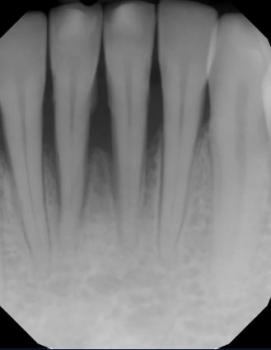
Patients with lower front gum inflammation may also experience tartar buildup, periodontal disease, gum abscesses, or tooth decay. Explore these related clinical case studies.