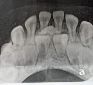
Why Are My Teeth Crumbling? A Severe Dental Decay Case Analysis
Severity:
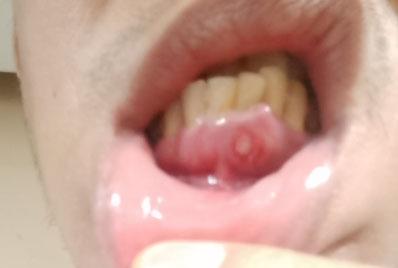
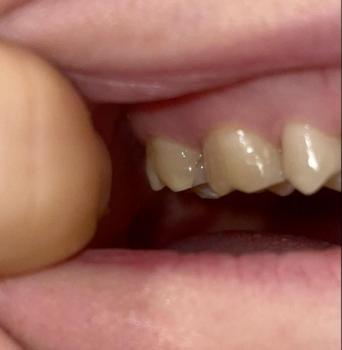
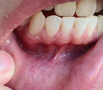
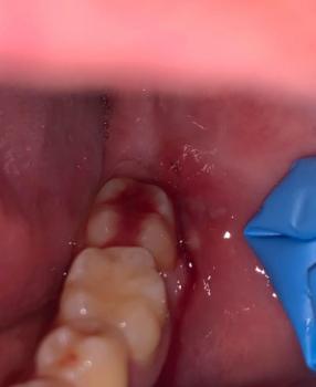
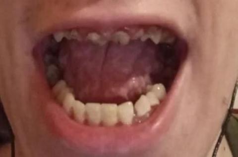
Teeth Problems:
Dental decay remains one of the most common chronic diseases worldwide, affecting millions of adults every year. While many cavities begin as small areas of enamel damage, untreated decay can progress into severe destruction of multiple teeth, leading to pain, infection, tooth loss, and significant impacts on quality of life.