Your Smile, Perfected with Precision.

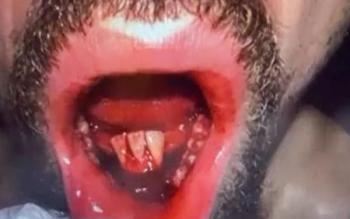
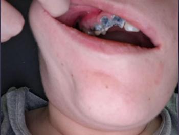
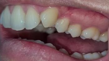
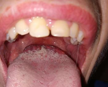
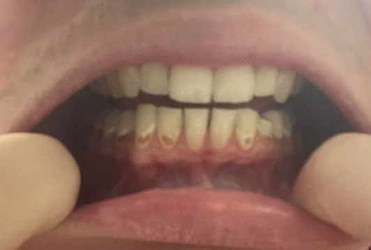
Early Enamel Erosion and Gum Inflammation in Lower Front Teeth - Zoomed Dental Case Analysis
Image:
Severity:
Teeth Problems:
Early Enamel Erosion on Lower Front Teeth: Can Dental Scaling Help?
Understanding the Difference Between Enamel Erosion, Tartar, and Tooth Decay
Discoloration or surface changes on the lower front teeth near the gumline can have several causes. In clinical practice, it is important to distinguish early enamel erosion from hardened dental calculus (tartar), gingivitis, and cervical dental caries because each condition requires a different approach.
Dental enamel is the hard, protective outer layer of the tooth. When it is repeatedly exposed to acids from soft drinks, acidic foods, or stomach reflux, minerals can dissolve from its surface. This process is called dental erosion. Unlike early bacterial tooth decay, which may be arrested or remineralized under appropriate conditions, enamel that has physically worn away through erosion cannot naturally grow back.
The lower front teeth are also a common location for tartar accumulation, particularly on the tongue-facing surfaces near the openings of the salivary glands. As a result, a patient may have both calculus deposits and enamel wear in the same region.
Additional Clinical: Early Enamel Erosion on the Lower Front Teeth — What Scaling Can and Cannot Treat
Clinical Insight: Is the Problem Tartar, Enamel Erosion, or Both?
When a patient presents with changes along the lower front teeth, the dentist must distinguish between hardened dental deposits, gum inflammation, tooth-surface wear, and actual enamel loss. These conditions can occur together, but they require different management approaches.
The lower front teeth are a common location for tartar accumulation, particularly on the tongue-facing surfaces near the openings of the salivary glands. Hardened deposits can contribute to gum inflammation and bleeding. However, an enamel defect or a smooth, worn tooth surface should not automatically be diagnosed as tartar-related damage.
Dental scaling removes plaque and hardened tartar; it does not rebuild eroded enamel. If early erosion is suspected, the dentist should assess the tooth surfaces before and after cleaning and determine whether the appearance reflects mineral loss, deposits, staining, or another condition.
How a Dentist Evaluates Early Enamel Erosion
A comprehensive examination considers several findings:
-
Surface appearance: Eroded enamel may appear unusually smooth, glossy, or lacking its normal surface texture.
-
Changes in tooth shape: Advanced wear can produce thinning, shallow depressions, or rounded edges.
-
Sensitivity: Some patients experience discomfort with cold drinks, hot foods, or sweet substances, although early erosion can be painless.
-
Dietary exposure: Frequent soft drinks, energy drinks, acidic fruit juices, and repeated acidic snacking may contribute to erosion.
-
Medical history: Acid reflux, recurrent vomiting, and conditions that reduce saliva can increase the risk of acid-related tooth wear.
-
Brushing habits: Aggressive brushing, particularly after acid exposure, can contribute to surface wear when combined with other risk factors.
These observations help guide the diagnosis, but appearance alone cannot reliably establish the severity or cause of enamel loss. A dentist may document the affected surfaces and compare them at future visits to assess progression.
Why Scaling Does Not Repair Enamel
Scaling is a preventive and periodontal procedure used to remove plaque and calculus from tooth surfaces, including deposits around or beneath the gumline when indicated.
If a patient has both tartar buildup and enamel erosion, scaling may improve gum health while leaving the underlying enamel defect unchanged. A tooth that appears rough or discoloured after cleaning does not necessarily mean the scaling procedure damaged the enamel; the cleaned surface may simply reveal a pre-existing defect.
Enamel that has been physically lost cannot naturally grow back. However, early mineral changes that have not yet resulted in permanent surface loss may sometimes be remineralized. The distinction is important because treatment depends on whether the problem is an early mineral imbalance, established erosion, decay, or another form of tooth wear.
Practical Strategies to Protect Remaining Enamel
1. Reduce frequent acid exposure
Reduce the frequency of acidic beverages, including soft drinks, energy drinks, and fruit juices. Avoid sipping them over several hours or repeatedly holding them in the mouth. Drinking water between meals and rinsing with water after acid exposure can help reduce the duration of contact.
2. Use fluoride toothpaste
Brush twice daily with fluoride toothpaste and a soft-bristled toothbrush. Use gentle pressure and avoid abrasive whitening products if your dentist identifies significant tooth wear. Fluoride can help protect teeth and support remineralization, although it cannot replace enamel that has already been lost.
3. Avoid brushing immediately after acid exposure
After consuming acidic drinks or foods, rinse with water and allow saliva time to neutralize the acidic environment before brushing. A commonly recommended precaution is to wait approximately 30–60 minutes, particularly after vomiting or significant acid exposure.
4. Investigate possible acid reflux
Repeated acid exposure from the stomach can damage teeth even when dietary habits are appropriate. If erosion is unexplained or progressing, discuss reflux symptoms or recurrent vomiting with a healthcare professional. Treating an underlying medical cause may be an important part of protecting the teeth.
5. Choose treatment according to the actual defect
Depending on the examination, management may involve preventive monitoring, fluoride-based measures, desensitizing products, or restorative treatment for significant structural loss. Small defects do not automatically require fillings or crowns. The objective is to control the cause, preserve remaining tooth structure, and restore function when necessary.
When Should You See a Dentist?
Arrange a dental assessment if you notice persistent sensitivity, thinning or chipping of the front teeth, changes in tooth shape, or gum bleeding that continues after cleaning.
Seek urgent dental or medical care if you develop facial swelling, fever with dental pain, or rapidly worsening symptoms. Difficulty swallowing or breathing requires emergency medical attention.
Clinical takeaway: The most appropriate treatment for lower-front-tooth changes depends on the diagnosis. Removing tartar may improve gum health, while protecting eroded enamel requires controlling acid exposure and monitoring any structural damage.
Frequently Asked Questions About Lower Front Tooth Enamel Erosion
1. Can dental scaling remove enamel erosion?
No. Scaling removes plaque and hardened tartar. It does not reverse or repair established enamel erosion. If erosion and tartar are both present, the dentist may treat the deposits and separately recommend measures to protect the remaining enamel.
2. Can scaling damage healthy enamel?
Professional scaling performed appropriately is intended to remove deposits while preserving tooth structure. Temporary sensitivity may occur, particularly when gums are inflamed or previously covered root surfaces become exposed. If you notice persistent pain or a new defect, have the area examined rather than assuming scaling caused enamel damage.
3. Why is tartar common behind the lower front teeth?
Saliva from nearby salivary gland openings contains minerals that can help plaque harden into calculus. This area can accumulate deposits even in people who brush regularly. Daily cleaning between the teeth and along the gumline helps control plaque, but hardened calculus generally requires professional removal.
4. Can early enamel erosion be reversed naturally?
Early mineral softening may be partly remineralized under favourable conditions, including adequate saliva and fluoride exposure. However, once enamel has been physically worn away, the lost structure does not regenerate naturally. A dentist can help determine which situation is more likely.
5. Does drinking soda cause enamel erosion?
Frequent exposure to acidic soft drinks can contribute to enamel erosion, including sugar-free varieties. The risk depends on factors such as acidity, frequency of consumption, contact time, and saliva. Reducing how often you drink them is generally more useful than simply switching to a sugar-free version.
6. Can enamel erosion cause sensitive lower front teeth?
Yes. As protective enamel becomes thinner, the underlying dentin may become exposed, making teeth more sensitive. However, sensitivity can also result from gum recession, tooth decay, cracks, or other dental problems, so a proper examination is necessary.
7. What toothpaste should I use if my enamel is eroding?
A fluoride toothpaste is a sensible starting point for most patients. Choose a product appropriate for your dental needs and avoid highly abrasive products if advised by your dentist. Special anti-erosion or desensitizing toothpastes may be helpful in selected cases, but no toothpaste can regrow lost enamel.
8. Can gum recession make enamel erosion look worse?
Yes. Gum recession exposes the root surface, which is covered by cementum and dentin rather than enamel. The exposed area may appear yellowish and become sensitive. A dentist should distinguish root exposure from enamel erosion because the prevention and treatment approaches differ.
9. Do I need a filling for early enamel erosion?
Not necessarily. Mild, stable erosion may only require preventive care and monitoring. Restorations may be considered when tooth structure loss causes persistent sensitivity, compromises function, creates significant aesthetic concerns, or continues to progress despite control of the underlying causes.
10. How often should enamel erosion be checked?
The appropriate interval depends on the severity, suspected cause, and evidence of progression. Your dentist may recommend shorter follow-up intervals for active or higher-risk erosion and longer intervals when the condition is stable. Photographs or clinical records can help identify changes over time.
11. Is enamel erosion the same as tooth decay?
No. Erosion is caused by direct chemical dissolution from acids that are not produced by oral bacteria. Tooth decay involves bacterial activity within dental plaque and the resulting acid attack. Both can damage teeth, and they can occur together.
12. Can enamel erosion be treated without crowns?
Often, yes. Early cases may be managed with preventive measures and monitoring. More substantial defects may be treated with appropriate adhesive restorations or other conservative options. Crowns are not automatically necessary; treatment should match the amount of remaining tooth structure and the patient's clinical needs.
Medical disclaimer: This educational content explains general dental principles and does not establish a diagnosis for an individual patient. A licensed dentist must examine the teeth and gums before recommending treatment.
A visual examination alone may not reliably distinguish all these conditions. A dentist should assess the tooth surface, gumline, plaque and calculus deposits, sensitivity, and possible signs of decay before recommending treatment.
1. What Does Dental Scaling Actually Treat?
Professional dental scaling removes hardened plaque deposits known as calculus, together with plaque and bacterial deposits from accessible tooth surfaces. When gum inflammation is caused by plaque accumulation, professional cleaning and improved home care can help reduce redness, bleeding, and swelling.
However, scaling does not rebuild lost enamel, reverse established erosive tooth wear, or automatically treat a cavity.
After tartar is removed, a previously covered area may look different. The natural tooth surface, existing discoloration, exposed root surface, or an underlying cavity may become more visible. This does not necessarily mean that scaling damaged the tooth.
Clinical distinction:
-
Tartar accumulation: Requires professional removal.
-
Plaque-induced gingivitis: Usually improves with professional cleaning and consistent oral hygiene.
-
Early enamel mineral loss: May benefit from fluoride and preventive measures, depending on the cause and whether actual tissue loss has occurred.
-
Established enamel erosion: Requires prevention of further acid exposure and monitoring; lost tooth structure cannot regenerate naturally.
-
Cavitated tooth decay: May require a restoration such as a filling.
2. How Can a Dentist Recognize Early Enamel Erosion?
Early erosive tooth wear may produce subtle changes that are difficult for patients to recognize without magnification and a proper dental examination.
Possible clinical findings include:
-
A smooth, unusually glossy, or softened-looking enamel surface.
-
Shallow depressions or loss of the natural tooth contours.
-
Increasing yellow appearance as enamel becomes thinner and the underlying dentin becomes more visible.
-
Sensitivity to cold, hot, or acidic foods and beverages.
-
Changes in the shape or appearance of the tooth edges.
-
Enamel loss that does not correspond to the usual pattern of bacterial cavities.
These findings are not exclusive to erosion. Yellow or brown areas near the gumline can also represent calculus, staining, exposed root dentin, or dental caries. The dentist must determine the most likely cause before treatment.
3. Why Does Enamel Erosion Develop?
Dental erosion occurs when acids chemically dissolve the mineral structure of teeth. Important risk factors include:
Frequent acidic drinks: Soft drinks, energy drinks, sports drinks, and many fruit juices can expose enamel to repeated acid attacks. Regularly sipping these beverages over several hours may increase the duration of acid exposure.
Acidic foods: Frequent consumption of highly acidic foods or snacks can contribute to erosive tooth wear, particularly when exposure is prolonged.
Gastroesophageal reflux: Stomach acid that repeatedly reaches the mouth can erode teeth. Recurrent vomiting can also cause substantial acid exposure.
Reduced saliva protection: Saliva helps neutralize acids and supports the natural repair of early mineral loss. Dry mouth can reduce this protective effect.
Aggressive brushing after acid exposure: Acid can temporarily soften the tooth surface. Brushing immediately afterward with excessive pressure or abrasive toothpaste may increase mechanical wear.
More than one factor may be involved. For example, a patient who frequently drinks soda and brushes aggressively may experience both chemical erosion and mechanical wear.
4. Recommended Dental Management for Early Enamel Erosion
Treatment should be based on the dentist's findings rather than on discoloration alone.
Step 1: Clinical examination
The dentist evaluates the lower front teeth, gumline, calculus deposits, enamel surface, and signs of cavities. Dental radiographs may be appropriate if decay or other disease is suspected, although X-rays do not directly measure early surface erosion.
Step 2: Professional scaling when indicated
If hardened calculus or plaque accumulation is present, professional scaling can improve oral hygiene and help control plaque-related gum inflammation. Polishing may be performed when clinically appropriate, but unnecessary or aggressive polishing should be avoided on vulnerable tooth surfaces.
Step 3: Fluoride-based prevention
A fluoride toothpaste can help protect against dental caries and support remineralization of early mineral loss. Depending on the patient's risk factors and examination, the dentist may recommend professional fluoride varnish or a prescription-strength fluoride product.
Fluoride does not replace enamel that has already been physically lost. Its role is preventive and protective, with benefits depending on the condition being treated.
Step 4: Reduce acid exposure
Patients should reduce the frequency of acidic drinks and avoid repeatedly sipping them throughout the day. Drinking water after acidic foods or beverages can help clear and dilute residual acids. When practical, consume acidic drinks with meals rather than between meals.
If reflux is suspected, medical assessment may be necessary to address the underlying cause.
Step 5: Review brushing technique
Brush twice daily for approximately two minutes using a soft-bristled toothbrush and fluoride toothpaste. Use gentle pressure and avoid aggressive scrubbing along the gumline. After consuming acidic food or drinks, rinse with water and wait approximately 30–60 minutes before brushing.
Step 6: Restore damaged teeth only when necessary
If examination confirms a cavity or clinically significant loss of tooth structure, the dentist may recommend a suitable restorative treatment. Small defects may be treated with composite resin, while more extensive damage may require other restorative options. Not every early erosion case needs a filling, veneer, or crown.
5. What Is a Realistic Recovery Timeline?
The timeline depends on whether the main problem is gum inflammation, early mineral loss, or permanent loss of tooth structure.
-
First few days after scaling: Mild gum tenderness or sensitivity can occur. Plaque-related inflammation may begin to improve.
-
Within one to two weeks: Gum bleeding and redness may decrease when professional cleaning is combined with effective daily oral hygiene.
-
Following weeks and months: The dentist can monitor whether preventive measures are controlling further erosive wear.
-
Long term: Continued protection depends on reducing acid exposure, maintaining oral hygiene, and attending appropriate dental reviews.
These are general expectations, not guaranteed healing deadlines. Enamel that has been physically lost does not grow back during a 7-day or 14-day recovery period. Persistent or worsening sensitivity requires reassessment.
6. When Should You Seek Further Dental Care?
Arrange a dental appointment if you notice progressive yellowing, visible grooves or pits, recurring sensitivity, gum bleeding, or a suspected cavity near the gumline.
Seek urgent dental assessment for severe or increasing tooth pain, localized pus, facial swelling, or fever associated with a dental problem. Difficulty swallowing or breathing, or rapidly spreading swelling of the face or neck, requires emergency medical care.
Expert Dentist's Clinical Perspective
When evaluating possible early enamel erosion on the lower anterior teeth, the priority is to identify the cause of the surface change before choosing treatment. Calculus removal can improve gum health, but it must not be presented as a treatment that regenerates lost enamel.
The most effective preventive strategy combines accurate diagnosis, appropriate professional cleaning, fluoride-based protection when indicated, gentle brushing, reduced exposure to dietary acids, and investigation of possible gastric reflux. If the affected area is actually a cavity or an exposed root surface, the treatment plan must be adjusted accordingly.
The objective is to protect the remaining tooth structure, control contributing factors, preserve function, and prevent avoidable progression.
Clinical note: This article provides general educational information. A photograph or online description cannot establish a definitive diagnosis. Individual treatment recommendations require assessment by a licensed dental professional.
Frequently Asked Questions (FAQ)
1. Can dental scaling repair early enamel erosion on the lower front teeth?
No. Scaling removes hardened plaque and calculus but cannot replace enamel lost through acid erosion. If the surface has only early mineral changes without substantial tissue loss, fluoride and improved preventive care may help protect the remaining enamel.
2. Can scaling make my lower front teeth look more yellow?
Yes, the teeth may appear different after cleaning because calculus and surface deposits have been removed. This can reveal the natural color of the tooth, exposed dentin, or an existing defect. Yellowing after scaling does not automatically mean that the procedure damaged the enamel.
3. Is enamel erosion the same as a cavity?
No. Erosion is chemical loss of tooth minerals caused by acids that are not produced by oral bacteria. Dental caries involves bacterial activity and can progress to form a cavity. Both conditions can coexist, so the correct diagnosis matters.
4. Can early enamel erosion be reversed naturally?
Early mineral loss may be partly remineralized under suitable conditions, especially when the tooth surface remains intact. However, once enamel has been physically worn away, it cannot regenerate naturally. Prevention aims to stop or slow further damage.
5. Does drinking Coca-Cola contribute to enamel erosion?
Yes. Coca-Cola and other soft drinks contain acids that can contribute to enamel erosion when exposure is frequent. Sugar-containing varieties also increase the risk of tooth decay. Reducing the frequency of consumption and choosing water more often can help lower these risks.
6. Should I brush immediately after drinking soda?
It is better to rinse with water and wait approximately 30–60 minutes before brushing. This helps avoid brushing while acid-softened tooth surfaces may be more vulnerable to mechanical wear.
7. Can fluoride toothpaste stop enamel erosion?
Fluoride toothpaste helps prevent cavities and supports remineralization of early mineral loss. It may improve resistance to acid challenges, but it cannot rebuild enamel that has already been physically removed. Reducing acid exposure remains essential.
8. Why do my gums bleed around the lower front teeth?
Bleeding commonly occurs with plaque-induced gingivitis, especially where plaque and tartar accumulate near the gumline. Other causes are possible, so persistent bleeding should be assessed by a dentist. Professional cleaning, gentle brushing, and daily cleaning between the teeth can help control plaque-related inflammation.
9. Does everyone with enamel erosion need a filling?
No. A filling may be appropriate if there is a cavity or a defect requiring restoration. Early erosion without significant structural damage may be managed through prevention and monitoring. Treatment should be based on clinical findings rather than appearance alone.
10. How often should I have dental scaling?
The appropriate interval depends on your tartar accumulation, gum health, periodontal history, and individual risk factors. Some patients benefit from regular six-month reviews, while others need more frequent professional maintenance. Your dentist should recommend an interval based on your examination.
11. Can enamel erosion cause tooth sensitivity?
Yes. As enamel thins, the underlying dentin may become exposed, allowing temperature changes and other stimuli to trigger sensitivity. Sensitivity can also result from gum recession, cavities, cracks, or other conditions, so persistent symptoms should be evaluated.
12. When is enamel erosion considered serious?
Erosion becomes more concerning when it progresses, exposes dentin, causes persistent sensitivity, changes the shape of teeth, or compromises their strength and function. Early evaluation is valuable because preventive treatment is generally simpler than restoring substantial lost tooth structure.
13. Can a dentist tell the difference between tartar and enamel erosion from a photograph?
A photograph can show suspicious discoloration, deposits, or surface changes, but it cannot reliably establish the diagnosis. A dentist may need direct examination, cleaning assessment, and additional tests to distinguish calculus, erosion, exposed dentin, and tooth decay.
14. Does enamel erosion always require veneers or crowns?
No. Many early cases do not require cosmetic or extensive restorative treatment. The priority is to control the cause and preserve the remaining tooth structure. Restorations are considered when damage, sensitivity, appearance, or function justifies them.
15. Can gum inflammation and enamel erosion occur at the same time?
Yes. Acid exposure can damage enamel while plaque and calculus cause gum inflammation. These conditions need separate but coordinated management: professional cleaning for plaque and calculus, preventive care for erosion, and restorative treatment if a cavity or substantial structural defect is present.
Suggested Internal Links
To strengthen the article's topical relevance, link naturally to related resources on your website where the content is available:
-
Gingivitis and bleeding gums: Explain plaque-related inflammation and professional cleaning.
-
Dental cavity treatment: Explain how dentists distinguish erosion from bacterial tooth decay.
-
Fluoride treatment: Discuss remineralization, prevention, and the limitations of fluoride.
-
Dental clinic directory in the Philippines: Help readers locate a licensed dentist for an examination.
Suggested anchor text: “learn more about bleeding gums,” “understand dental cavity treatment,” and “find a dental clinic near you.”
Suggested SEO Additions
SEO title: Early Enamel Erosion on Lower Front Teeth: Does Scaling Help?
Meta description: Learn how dentists distinguish enamel erosion, tartar, and cavities on lower front teeth, when scaling helps, and how fluoride and diet changes protect enamel.
Suggested FAQ schema topics: Dental scaling and enamel erosion; whether enamel can grow back; fluoride for erosion; soda and enamel damage; yellow teeth after scaling; and when a filling is needed.
Editorial recommendation: Keep the original case observations clearly labeled as image-based findings. Avoid presenting erosion, caries, or a precise healing timeline as confirmed without an in-person examination.
Teeth Case – Zoom 100%
Focused Area: Lower front teeth (anterior region) and gingival margin
Full Analysis and Deep Visual Examination
This assessment is based on the provided image only and does not replace a comprehensive dental examination, periodontal probing, or dental X-rays.
Observed Findings
-
Yellow-brown lesions on the lower front teeth near the gumline, consistent with early caries and enamel erosion
-
Gingival redness and inflammation along the lower anterior gum margin
-
Signs of acid-related enamel wear, commonly associated with frequent soda or acidic beverage intake
-
Plaque accumulation along the cervical (neck) area of the teeth
-
No obvious fractures, but surface integrity is compromised
Probable Diagnosis
-
Early dental caries of the lower anterior teeth
-
Acid erosion of enamel (diet-related)
-
Plaque-induced gingivitis
-
Increased risk of cervical decay progression if untreated
Recommended Treatment Process
Step 1: Full Mouth Scaling and Polishing (Day 1)
-
Ultrasonic scaling to remove plaque and calculus
-
Manual scaling along the gumline of the lower front teeth
-
Polishing to smooth enamel surfaces and reduce plaque retention
Estimated duration: 30–60 minutes
Local anesthesia: Usually not required unless sensitivity is present
Step 2: Enamel Protection and Caries Control (Days 1–7)
-
Fluoride varnish or remineralization treatment
-
Assessment for small composite fillings if lesions are cavitated
-
Dietary counseling to reduce acidic and sugary drink intake
Healing and Recovery Timeline
| Time Frame | Expected Outcome |
|---|---|
| Days 1–3 | Reduced gum redness and irritation |
| Days 4–7 | Gum tissue tightens; sensitivity decreases |
| Days 8–14 | Stable gum health if acids are avoided |
A 14-day healing period is realistic for gum tissue, provided acidic exposure is controlled.
What Will Scale Up If Left Untreated
-
Progression of enamel erosion into deeper dentin
-
Formation of visible cavities requiring fillings
-
Increased tooth sensitivity and pain
-
Worsening gingival inflammation
-
Higher risk of periodontal involvement in the lower front teeth
Post-Treatment Care Instructions
-
Brush twice daily using a soft-bristle toothbrush
-
Use high-fluoride toothpaste if recommended by a dentist
-
Floss daily, especially between lower front teeth
-
Rinse with water after consuming acidic drinks
-
Limit soda, energy drinks, and citrus beverages
Professional Comment
This case shows early but active damage, likely linked to dietary acids combined with plaque buildup. With timely scaling, fluoride therapy, and dietary changes, progression can be stopped and enamel integrity preserved.
Visit a Dental Clinic Near You
For professional scaling, fluoride treatment, and decay assessment, find a nearby dental clinic using our directory:
Video :
Related Clinical Cases
Patients with lower front gum inflammation may also experience tartar buildup, periodontal disease, gum abscesses, or tooth decay. Explore these related clinical case studies.